Skip directly to site content Skip directly to page options Skip directly to A-Z link Skip directly to A-Z link Skip directly to A-Z link
Volume 30, Number 7—July 2024
Research

Sialic Acid Receptor Specificity in Mammary Gland of Dairy Cattle Infected with Highly Pathogenic Avian Influenza A(H5N1) Virus

Rahul K. Nelli1Comments to Author , Tyler A. Harm1, Chris Siepker1, Jennifer M. Groeltz-Thrush, Brianna Jones, Ning-Chieh Twu, Ariel S. Nenninger, Drew R. Magstadt, Eric R. Burrough, Pablo E. Piñeyro, Marta Mainenti, Silvia Carnaccini, Paul J. Plummer, and Todd M. BellComments to Author 
Author affiliations: Iowa State University College of Veterinary Medicine, Ames, Iowa, USA (R.K. Nelli, T.A. Harm, C. Siepker, J.M. Groeltz-Thrush, B. Jones, N.-C. Twu, A.S. Nenninger, D.R. Magstadt, E.R. Burrough, P.E. Piñeyro, M. Mainenti, P.J. Plummer, T.M. Bell); University of Georgia, Athens, Georgia, USA (S. Carnaccini)

Main Article

Figure 2

Respiratory tract tissues from a US dairy cow infected with highly pathogenic avian influenza A(H5N1) virus, showing IAV-Np (teal chromogen), individually duplexed with MAL-I (magenta chromogen), MAL-II (magenta chromogen), and SNA (magenta chromogen) using chromogenic staining. Representative images are shown for IAV-Np/MAL-I (A, D, G, J), IAV-Np/MAL-II (B, E, H, K), and IAV-Np/SNA (C, F, I, L) are shown. No IAV-Np was observed in the unaffected respiratory tissue sections. Intense granular to punctate labeling for MAL-I (A) and MAL-II (B) were observed within goblet cells (arrowheads), along the apical ciliated margin (arrows), and glands of the trachea. SNA labeling (C) was confined to intraepithelial round cells (arrows), endothelium, and glands of the trachea. Similar labeling for MAL-I (D) and MALII (E) was observed within the bronchial lumen within goblet cells (arrowheads) and along the apical cell margin (arrows). SNA (F) labeling was only observed in rare goblet cells (arrowheads) and lamina proprial round cells (arrows) in the bronchus. Bronchioles had diffuse, fine, fibrillary to apical membranous labeling (arrows) MAL-I (G) and MALII (H). No substantial labeling was detected within the mucosal epithelial cells of bronchioles with SNA (I). Diffuse, fine, apical membranous labeling of pneumocytes lining alveoli was observed with MAL-I (J) and MAL-II (K) (arrows). SNA (L) labeling within the alveolar portions of the lung was confined to endothelium and interstitial round cells. Scale bars indicate 200 μm. IAV-Np, influenza A virus nucleoprotein; MAL, Maackia amurensis lectin; SNA, Sambucus nigra lectin.

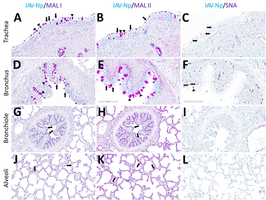
Figure 2. Respiratory tract tissues from a US dairy cow infected with highly pathogenic avian influenza A(H5N1) virus, showing IAV-Np (teal chromogen), individually duplexed with MAL-I (magenta chromogen), MAL-II (magenta chromogen), and SNA (magenta chromogen) using chromogenic staining. Representative images are shown for IAV-Np/MAL-I (A, D, G, J), IAV-Np/MAL-II (B, E, H, K), and IAV-Np/SNA (C, F, I, L) are shown. No IAV-Np was observed in the unaffected respiratory tissue sections. Intense granular to punctate labeling for MAL-I (A) and MAL-II (B) were observed within goblet cells (arrowheads), along the apical ciliated margin (arrows), and glands of the trachea. SNA labeling (C) was confined to intraepithelial round cells (arrows), endothelium, and glands of the trachea. Similar labeling for MAL-I (D) and MALII (E) was observed within the bronchial lumen within goblet cells (arrowheads) and along the apical cell margin (arrows). SNA (F) labeling was only observed in rare goblet cells (arrowheads) and lamina proprial round cells (arrows) in the bronchus. Bronchioles had diffuse, fine, fibrillary to apical membranous labeling (arrows) MAL-I (G) and MALII (H). No substantial labeling was detected within the mucosal epithelial cells of bronchioles with SNA (I). Diffuse, fine, apical membranous labeling of pneumocytes lining alveoli was observed with MAL-I (J) and MAL-II (K) (arrows). SNA (L) labeling within the alveolar portions of the lung was confined to endothelium and interstitial round cells. Scale bars indicate 200 μm. IAV-Np, influenza A virus nucleoprotein; MAL, Maackia amurensis lectin; SNA, Sambucus nigra lectin.

Main Article

1These first authors contributed equally to this article.

Page created: June 03, 2024
Page updated: June 22, 2024
Page reviewed: June 22, 2024
The conclusions, findings, and opinions expressed by authors contributing to this journal do not necessarily reflect the official position of the U.S. Department of Health and Human Services, the Public Health Service, the Centers for Disease Control and Prevention, or the authors' affiliated institutions. Use of trade names is for identification only and does not imply endorsement by any of the groups named above.
file_external