Volume 30, Number 7—July 2024
Research
Sialic Acid Receptor Specificity in Mammary Gland of Dairy Cattle Infected with Highly Pathogenic Avian Influenza A(H5N1) Virus
Figure 3
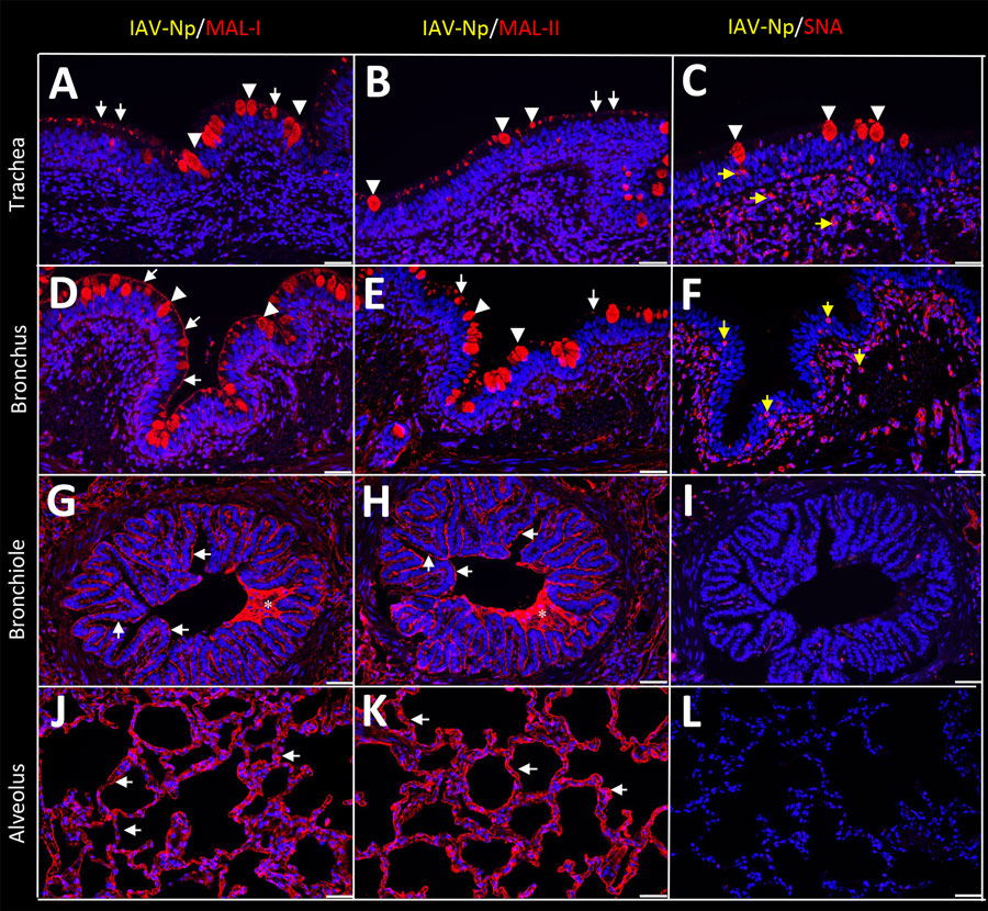
Figure 3. Respiratory tract tissues from a US dairy cow infected with highly pathogenic avian influenza A(H5N1) virus, labeled with IAV-Np (yellow pseudocolor, DyLight 594), individually duplexed with MAL-I (red pseudocolor, Alexa Fluor 647), MAL-II (red pseudocolor, Alexa Fluor 647), and SNA (red pseudocolor, Alexa Fluor 647) using fluorescent staining. Representative merged images are shown for IAV-Np and MAL-I (A, D, G, J), IAV-Np and MAL-II (B, E, H, K), and IAV-Np and SNA (C, F, I, L). IAV-Np labeling was not detected within the respiratory tissue sections. Intense, granular to punctate, cytoplasmic MAL-I, MAL-II, and SNA labeling was observed in goblet cells (arrowheads) and glands of the trachea (A–C). Similar goblet cell labeling (arrowheads) for MAL-I (D) and MAL-II (E) was observed in the bronchi with weak SNA labeling (F). Multifocal, moderate, fibrillar, apical, membranous MAL-I (A) and MAL-II (B) labeling (white arrows) was observed on the tracheal epithelium. The respiratory epithelium of the bronchi, bronchioles, and alveoli had diffuse, moderate to intense, apical, fibrillar MAL-I labeling (white arrows) (D, G, J). The respiratory epithelium of the bronchi had multifocal, moderate, fibrillar, apical MAL-II labeling (white arrows) (E). The respiratory epithelium of the bronchioles and alveoli had diffuse MAL-II labeling (white arrows) (H, K). Intraluminal secretory material (asterisks) in the bronchi and bronchioles were intensely labeled with MAL-I and MAL-II (G, H). Membranous, granular SNA labeling (yellow arrows) was observed in intraepithelial and lamina proprial round cells in the trachea and bronchi (C, F). Scale bars indicate 50 μm. IAV-Np, influenza A virus nucleoprotein; MAL, Maackia amurensis lectin; SNA, Sambucus nigra lectin.
1These first authors contributed equally to this article.