Skip directly to site content Skip directly to page options Skip directly to A-Z link Skip directly to A-Z link Skip directly to A-Z link

Disclaimer: Early release articles are not considered as final versions. Any changes will be reflected in the online version in the month the article is officially released.

Volume 30, Number 8—August 2024
Research

Scrapie Versus Chronic Wasting Disease in White-Tailed Deer

Zoe J. Lambert1, Jifeng Bian, Eric D. Cassmann, M. Heather West Greenlee, and Justin J. GreenleeComments to Author 
Author affiliations: Oak Ridge Institute for Science and Education, Oak Ridge, Tennessee, USA (Z.J. Lambert); US Department of Agriculture, Ames, Iowa, USA (Z.J. Lambert, J. Bian, E.D. Cassmann, J.J. Greenlee); Iowa State University, Ames (Z.J. Lambert, M.H. West Greenlee)

Main Article

Figure 1

Immunohistochemistry demonstrating misfolded prion protein in white-tailed deer oronasally inoculated with white-tailed deer scrapie (WTD scrapie) agent in study of scrapie versus chronic wasting disease (CWD) in white-tailed deer. A) Vacuolation in the dorsal motor nucleus of the vagus in the brain stem at the level of the obex of each deer. B–D) Misfolded prion protein in the dorsal motor nucleus of the vagus in the brain stem at the level of the obex (B), palatine tonsil (C), and retropharyngeal lymph node (D) of each deer. E–H) Neurotropism of the scrapie form of the prion protein for retinal ganglion cells with scrapie agent (closed arrowheads) and not CWD (open arrowhead). E) Sheep scrapie retina; F) WTD scrapie, passage 1, retina; G) WTD scrapie, passage 2, retina; H) WTD CWD, retina. Hematoxylin and eosin staining; original magnification ×10 for panels A–D, ×20 for panels E–H. GCL, ganglion cell layer; INL, inner nuclear layer; IPL, inner plexiform layer; ONL, outer nuclear layer; OPL, outer plexiform layer.

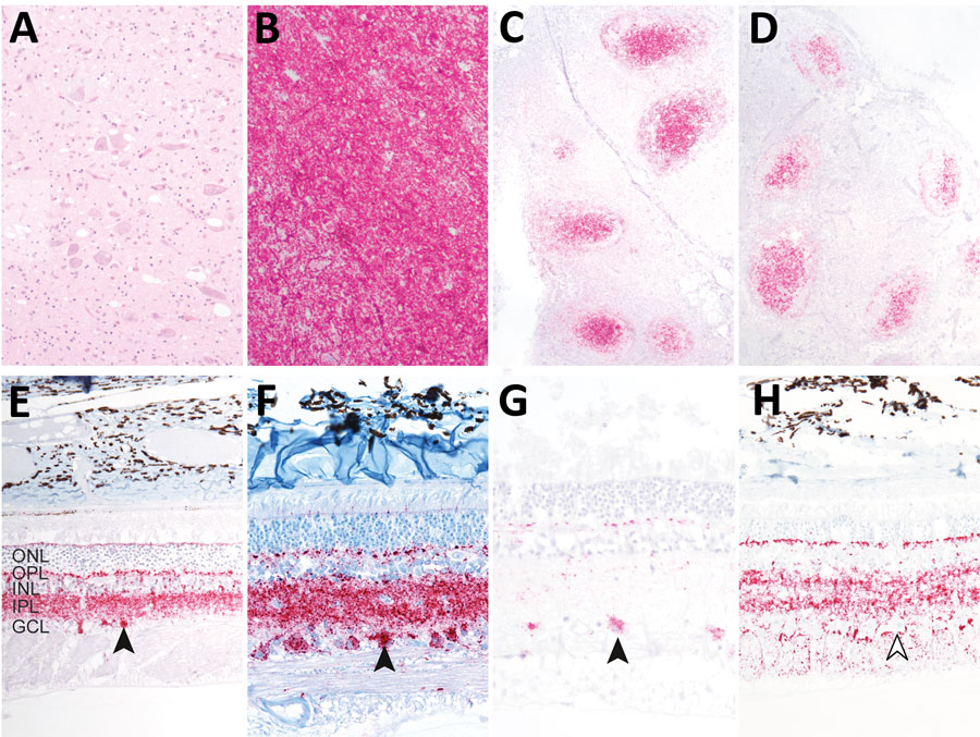
Figure 1. Immunohistochemistry demonstrating misfolded prion protein in white-tailed deer oronasally inoculated with white-tailed deer scrapie (WTD scrapie) agent in study of scrapie versus chronic wasting disease (CWD) in white-tailed deer. A) Vacuolation in the dorsal motor nucleus of the vagus in the brain stem at the level of the obex of each deer. B–D) Misfolded prion protein in the dorsal motor nucleus of the vagus in the brain stem at the level of the obex (B), palatine tonsil (C), and retropharyngeal lymph node (D) of each deer. E–H) Neurotropism of the scrapie form of the prion protein for retinal ganglion cells with scrapie agent (closed arrowheads) and not CWD (open arrowhead). E) Sheep scrapie retina; F) WTD scrapie, passage 1, retina; G) WTD scrapie, passage 2, retina; H) WTD CWD, retina. Hematoxylin and eosin staining; original magnification ×10 for panels A–D, ×20 for panels E–H. GCL, ganglion cell layer; INL, inner nuclear layer; IPL, inner plexiform layer; ONL, outer nuclear layer; OPL, outer plexiform layer.

Main Article

1Current affiliation: Des Moines University, Des Moines, Iowa, USA.

Page created: June 14, 2024
Page updated: July 16, 2024
Page reviewed: July 16, 2024
The conclusions, findings, and opinions expressed by authors contributing to this journal do not necessarily reflect the official position of the U.S. Department of Health and Human Services, the Public Health Service, the Centers for Disease Control and Prevention, or the authors' affiliated institutions. Use of trade names is for identification only and does not imply endorsement by any of the groups named above.
file_external