Volume 32, Number 5—May 2026
Synopsis
Borna Disease Virus 1 as Cause of Fatal Meningoencephalomyelitis in Wild Hedgehogs, Germany, 2022–2025
Figure 4
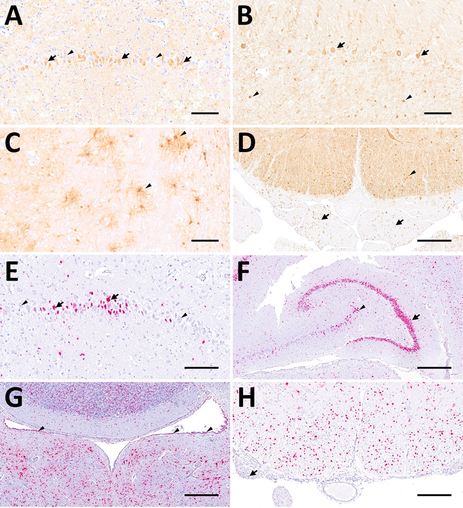
Figure 4. Distribution of Borna disease virus 1 detected within the CNS of infected hedgehogs in study of the virus as cause of fatal meningoencephalomyelitis in wild hedgehogs, Germany, 2022–2025. A–D) Antigen detected by immunohistochemistry by using Bo18 antibody. A) Multiple immunopositive neurons within the pyramidal layer of the hippocampus (arrows) mingling with negative neurons (arrowheads) from case 1. Scale bar represents 100 μm. B) Multiple immunopositive Purkinje (arrows) and few granule cells (arrowheads) within the cerebellar cortex from case 3. Scale bar represents 100 μm. C) Multiple positive astroglial cells within the spinal white matter (arrowheads) from case 6. Scale bar represents 100 μm. D) Multiple positive oligondendroglial cells within the spinal white matter (arrowhead), as well as multiple positive nerve fibers within the adjacent nerve roots (arrows) from case 5. Scale bar represents 250 μm. E–H) Virus RNA detected by RNAscope (Advanced Cell Diagnostics, Inc., https://acdbio.com) in situ hybridization (ISH). E) Numerous ISH-positive neurons (arrows) mingling with negative ones (arrowheads) within the corresponding area of the hippocampus shown in (A) from case 1. Scale bar represents 100 μm. F) Granule cells of dentate gyrus (arrow) are almost entirely ISH-positive as do multiple neurons of cornu ammonis (arrowhead) from case 5. Scale bar represents 500 μm. G) Numerous ISH-positive neurons, glial as well as ependymal cells (arrowheads) surrounding the fourth ventricle from case 7. Scale bar represents 500 μm. H) Numerous ISH- positive oligodendroglial cells within the spinal white matter and rare signals within nerve roots (arrow) from case 5. Scale bar represents 250 μm. Stains: A–I, 3,3′-diaminobenzidine with hematoxylin counterstain; E–H, 2.5 HD assay–RED with hematoxylin counterstain.
1These senior authors contributed equally to this article.