Borna Disease Virus 1 as Cause of Fatal Meningoencephalomyelitis in Wild Hedgehogs, Germany, 2022–2025
Effrosyni Michelakaki, Benjamin Schade, Brigitte Boehm, Eva Kappe, Marcel Suchowski, Anne Kupca, Magdalena Schumacher, Anna Maria Gager, Friederike Liesche-Starnecker, Sonja Fiedler, Eva Schwarz, Zoltan Bago, Andreas Blutke, Martin Beer, Dennis Rubbenstroth
1
, and Kaspar Matiasek
1
Author affiliation: Centre for Clinical Veterinary Medicine, Ludwig Maximilians-Universität München, Munich, Germany (E. Michelakaki, S. Fiedler, E. Schwarz, A. Blutke, K. Matiasek); Bavarian Animal Health Service, Poing, Germany (B. Schade, B. Boehm, E. Kappe); Bavarian Health and Food Safety Authority, Oberschleißheim, Germany (M. Suchowski, A. Kupca, M. Schumacher, A.M. Gager); University Medical Center Ulm, Ulm University, Ulm, Germany (F. Liesche-Starnecker); Institute for Veterinary Disease Control, Mödling, Austria (Z. Bago); Institute of Diagnostic Virology, Friedrich-Loeffler-Institut, Greifswald-Insel Riems, Germany (M. Beer, D. Rubbenstroth)
Main Article
Figure 7
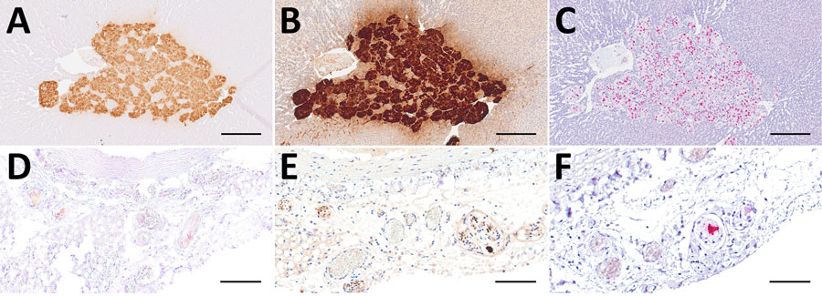
Figure 7. Comparison of 3 diagnostic methods, in the adrenal glands (A–C) and mediastinal nerve branches (D–F) of case 5, a hedgehog from Germany with Borna disease virus 1 (BoDV-1) infection, in study of the virus as cause of fatal meningoencephalomyelitis in wild hedgehogs, Germany, 2022–2025. Diffusely positive chromaffin cells and multifocal positive nerve branches are seen by immunohistochemistry by using BoDV-1 nucleoprotein mouse monoclonal antibody Bo18 (A, D), rabbit anti–BoDV-1 nucleoprotein polyclonal hyperimmune serum #201 (B, E) and RNAscope (Advanced Cell Diagnostics, Inc., https://acdbio.com) in situ hybridization (C, F). Scale bars: A–C, 250 μm; D–F, 100 μm. Stains: A, B, D, E, 3,3′-diaminobenzidine with hematoxylin counterstain; C, F, 2.5 HD assay–RED with hematoxylin counterstain.
Main Article
Page created: April 02, 2026
Page updated: May 06, 2026
Page reviewed: May 06, 2026
The conclusions, findings, and opinions expressed by authors contributing to this journal do not necessarily reflect the official position of the U.S. Department of Health and Human Services, the Public Health Service, the Centers for Disease Control and Prevention, or the authors' affiliated institutions. Use of trade names is for identification only and does not imply endorsement by any of the groups named above.