Volume 32, Number 5—May 2026
Dispatch
Development and Validation of Real-Time PCR for Detecting Anaplasma bovis–Like Agent in Dermacentor spp. Ticks
Cite This Article
Citation for Media
Abstract
We developed and validated a real-time PCR to detect an Anaplasma bovis–like agent. We applied that assay to 672 Dermacentor spp. ticks collected from across the United States and found 0.1% A. bovis–like agent prevalence. This assay could enhance epidemiologic surveys for this A. bovis–like agent in ticks and humans.
An Anaplasma bovis–like agent was detected in humans in the United States in 2020 and was further described from 4 patients and a few Dermacentor spp. ticks in 2023 (1,2). Clinical data from human cases was not available, although patient samples were submitted for suspicion of tickborne illness (1,2). A subsequent regional survey detected the A. bovis–like agent in 10.1% (4/38) of D. variabilis ticks from Oklahoma but not in 93 Kansas D. variabilis ticks or 140 Oklahoma beef cattle (3).
Human illness caused by A. bovis has been described in China, where infection of ruminants is common (4,5). Epidemiologic investigation of the A. bovis–like agent is obstructed by lack of a specific, rapid molecular assay. Previous studies have relied on conventional PCRs (2,3), which are suitable for genetic characterization but cumbersome for screening large sample sets. To improve diagnostic methods, we developed and optimized a real-time probe-based PCR assay specifically for the A. bovis–like agent.
We retrieved from GenBank heat shock chaperon gene (groEL) sequences from the A. bovis–like agent and related Anaplasma spp. bacteria, including A. bovis, A. capra, A. centrale, A. marginale, A. ovis, A. phagocytophilum, and A. platys (Appendix Table). We aligned sequences and generated potential primers and probes in Geneious Prime version 2025.2.2 (https://www.geneious.com) by using the primer design tool. We generated 1 assay (AboviTM) targeting a 125-bp fragment of the groEL gene. AboviTM satisfied the design parameters, and we expected it to be specific to A. bovis–like agent because of base mismatches with nontarget sequences within the oligo binding sites.
We had custom AboviTM oligos synthesized in TaqMan assays (Thermo Fisher Scientific, https://www.thermofisher.com), then further interrogated the oligos (Table 1). We performed PCR reactions in MicroAmp EnduraPlate Optical 96-Well Clear Reaction Plates on an Applied Biosystems QuantStudio 3 Real-Time PCR instrument (both Thermo Fisher Scientific) and included nontemplate controls for every run.
We initially verified AboviTM by amplifying (×3) four A. bovis–like DNA samples previously extracted from D. variabilis ticks, then confirming by conventional PCR (3). Each 20-μL PCR reaction contained 10 μL TaqMan Fast Advanced Master Mix (Thermo Fisher Scientific), 900 nmol of each primer, 250 nmol probe, 2 μL DNA template, and molecular grade water. Thermocycling conditions were enzyme activation at 95°C for 60 seconds, followed by 45× cycles of denaturation at 95°C for 3 seconds, and annealing/extension at 60°C for 30 seconds. To confirm appropriate amplicon size and identity, we performed gel electrophoresis in 2.0% agarose gel, followed by PCR product purification using the Wizard SV Gel and PCR Clean-up Kit (Promega, https://www.promega.com). Oklahoma State University (OSU) Molecular Core Facility performed Sanger sequencing (unidirectional), and we confirmed sequence identity by BLAST analysis (https://blast.ncbi.nlm.nih.gov).
For further assay optimization, we used a gBlocks synthetic double-stranded DNA standard (Integrated DNA Technologies, https://www.idtdna.com) of the target sequence. We prepared a series of 10-fold dilutions to attain a range of 1–100,000,000 copies of target DNA per reaction. We evaluated combinations of varying primer (75–900 nmol) and probe (100–250 nmol) concentrations by using 100 copies/reaction. We considered assay performance to be optimal when the amplification curve demonstrated concurrently high baseline-corrected normalized reporter and low cycle threshold value (6). Assay performance was optimal at primer concentrations of 900 nmol each and probe concentration of 250 nmol. We performed subsequent PCR reactions with those concentrations and the thermocycling protocol described above.
We conducted standard curve analyses to evaluate the AboviTM assay efficiency and sensitivity by using 2 sample sets: the 10-fold dilution series containing synthetic target DNA only and the same dilution series with ≈54 ng of noninfected D. variabilis tick DNA added per reaction. We used tick samples to assess assay performance in the tick DNA matrix and obtained noninfected adult ticks from the OSU Tick Rearing Facility.
Figure 1

Figure 1. Results of the standard curve analyses, correlation, and amplification efficiency of real-time PCR for detecting Anaplasma bovis–like agent in Dermacentorspp. ticks, United States. We conducted standard...
We amplified both dilution series in triplicate and repeated that experiment over 5 independent runs. We checked raw PCR data for quality, then used Applied Biosystems Design & Analysis Software version 2.8.0 (Thermo Fisher Scientific) to analyze data and exported data into Excel Version 1808 (Microsoft, https://www.microsoft.com). We calculated standard curve regression, amplification efficiency, and correlation by using the qPCRtools package in R Studio 2024.09.1 (7). We used the ggplot2 package in R Studio to visualize statistical and cycle threshold data (Figure 1).
The assay’s limit of detection was 10 target copies. AboviTM detected as low as 1 target copy; however, limit of detection <3 copies is considered invalid because of stochastic limitations of quantitative PCR (8). We interrogated specificity by running AboviTM in triplicate with genetically similar agents that might be detected in ticks or mammals and observed no cross-amplification against Anaplasma phagocytophilum, A. marginale, A. platys, Ehrlichia ewingii, E. chaffeensis, Rickettsia bellii, or R. montanensis. Those nontarget DNA samples were previously extracted from ticks or mammalian whole blood and confirmed by PCR and Sanger sequencing (data not shown).
Figure 2
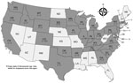
Figure 2. Number and geographic distribution of Dermacentor spp. ticks collected from dogs and cats and tested by real-time PCR (AboviTM assay) for Anaplasma bovis–like agent, United States....
We then applied the optimized AboviTM assay to 672 Dermacentor spp. ticks collected from dogs and cats across the United States during 2021–2023 (Figure 2). Veterinary clinics shipped ticks to the OSU College of Veterinary Medicine (https://www.showusyourticks.org), where staff morphologically identified ticks and stored in 70% ethanol at −20°C until dissection and DNA extraction.
We performed dissection by adhering ticks, ventral side down, on glass slides using double-sided adhesive tape. We used sterile no. 11 scalpels to open each tick along the coronal plane and to scrape out internal contents, which we transferred to sterile 1.5 mL microfuge tubes. We discarded tick exoskeletons. We used Cytiva Blood Genomic Prep Mini Spin Kit (Cytiva, https://www.cytivalifesciences.com) to extract total genomic DNA from tick samples, according to manufacturer recommendations. We tested <1 female and <1 male tick per species from an individual host. We detected A. bovis–like agent from only 1 (0.1%) tick (Table 2): a male D. variabilis tick collected from a dog in Blaine, Minnesota. We further verified that positive sample by using conventional PCRs targeting the 16S (rrs), citrate synthetase (gltA), and groEL genes, as previously described (2,9,10). We resolved amplicons by gel electrophoresis, then purified, performed Sanger sequencing (unidirectional), and analyzed as described. All sequences were 100% identical to previously reported sequences from Dermacentor spp. ticks and humans in the United States (GenBank accession nos. OQ772255, OQ772257.1, OQ693619, and PQ166304–7).
Few cases of human A. bovis–like infection have been reported in the United States (1,2). Some data suggest Dermacentor spp. tick transmission, although we detected A. bovis–like DNA in only 0.1% of Dermacentor spp. ticks tested. In our previous survey, we detected A. bovis–like DNA in 10.1% (4/38) of D. variabilis ticks from Oklahoma, 3 (75%) of which originated from the same collection site (3). Distribution of A. bovis might be highly focalized because, including findings from this study, A. bovis–like DNA has only been detected in humans and D. variabilis ticks from central US states, including Minnesota, Oklahoma, Missouri, Iowa, and Nebraska, and in D. andersoni ticks from Saskatchewan, Canada (2,3). Other possible tick and mammal species involved in transmission are currently unknown.
Because A. bovis–like agent has been detected in few Dermacentor spp. ticks and humans and few studies have investigated its epidemiology, broad conclusions cannot yet be drawn about its complete geographic distribution. AboviTM enables rapid, specific detection for A. bovis–like agent with high efficiency, dramatically improving methods for screening samples. Application of AboviTM to future studies could enable further largescale screening of humans, potential vectors, and animal hosts, which could greatly improve epidemiologic understanding of A. bovis–like agent in the United States.
Dr. Smith is a recent graduate of the Oklahoma State College of Veterinary Medicine Comparative Biomedical Sciences graduate program. Her research interests include epidemiology and diagnosis of emerging vectorborne infections of zoonotic and veterinary importance and clinical diagnostic parasitology.
R.C.S. designed the study, performed and supervised most of the laboratory work described, wrote the first draft of the manuscript, and was responsible for funding acquisition. A.C. and D.F.B.M. performed DNA extraction from tick specimens. K.T.D. provided insight on experimental design of qPCR optimization and instrument use. L.A.S. assisted with funding acquisition. All authors reviewed and approved the final version of this article.
Acknowledgment
We thank Susan Little, Kellee Sundstrom, and all other past and present OSU College of Veterinary Medicine personnel who have supported the Show Us Your Ticks Project, the source of ticks used for this study. We also thank Wade Burton and Ruth Scimeca for providing DNA-positive controls for A. platys and A. marginale, respectively. We thank the National Center for Veterinary Parasitology, who provided research grant funding in support of this study (primary investigator R.C.S.).
References
- Kingry L, Sheldon S, Oatman S, Pritt B, Anacker M, Bjork J, et al. Targeted metagenomics for clinical detection and discovery of bacterial tick-borne pathogens. J Clin Microbiol. 2020;58:e00147–20. DOIPubMedGoogle Scholar
- Karpathy SE, Kingry L, Pritt BS, Berry JC, Chilton NB, Dergousoff SJ, et al. Anaplasma bovis–like infections in humans, United States, 2015–2017. Emerg Infect Dis. 2023;29:1904–7. DOIPubMedGoogle Scholar
- Smith RC, Myers S, Sundstrom KD, Wilson R, Scimeca RC, Starkey LA, et al. Detection of Anaplasma bovis–like agent in the southcentral United States. Ticks Tick Borne Dis. 2024;15:
102411 . DOIPubMedGoogle Scholar - Lu M, Chen Q, Qin X, Lyu Y, Teng Z, Li K, et al. Outbreak reports: Anaplasma bovis infection in fever and thrombocytopenia patients—Anhui Province, China, 2021. China CDC Wkly. 2022;4:249–53. DOIPubMedGoogle Scholar
- Liu Z, Ma M, Wang Z, Wang J, Peng Y, Li Y, et al. Molecular survey and genetic identification of Anaplasma species in goats from central and southern China. Appl Environ Microbiol. 2012;78:464–70. DOIPubMedGoogle Scholar
- Raymaekers M, Smets R, Maes B, Cartuyvels R. Checklist for optimization and validation of real-time PCR assays. J Clin Lab Anal. 2009;23:145–51. DOIPubMedGoogle Scholar
- Li X, Wang Y, Li J, Mei X, Liu Y, Huang H. qPCRtools: an R package for qPCR data processing and visualization. Front Genet. 2022;13:
1002704 . DOIPubMedGoogle Scholar - Bustin SA, Benes V, Garson JA, Hellemans J, Huggett J, Kubista M, et al. The MIQE guidelines: minimum information for publication of quantitative real-time PCR experiments. Clin Chem. 2009;55:611–22. DOIPubMedGoogle Scholar
- Sumner JW, Nicholson WL, Massung RF. PCR amplification and comparison of nucleotide sequences from the groESL heat shock operon of Ehrlichia species. J Clin Microbiol. 1997;35:2087–92. DOIPubMedGoogle Scholar
- Zhuang L, Du J, Cui XM, Li H, Tang F, Zhang PH, et al. Identification of tick-borne pathogen diversity by metagenomic analysis in Haemaphysalis longicornis from Xinyang, China. Infect Dis Poverty. 2018;7:45. DOIPubMedGoogle Scholar
Figures
Tables
Cite This ArticleOriginal Publication Date: April 28, 2026
Table of Contents – Volume 32, Number 5—May 2026
| EID Search Options |
|---|
|
|
|
|
|
|
Please use the form below to submit correspondence to the authors or contact them at the following address:
Rachel C. Smith, Oklahoma State University College of Veterinary Medicine, 250 McElroy Hall, 208 N McFarland St, Stillwater, OK 74078, USA
Top