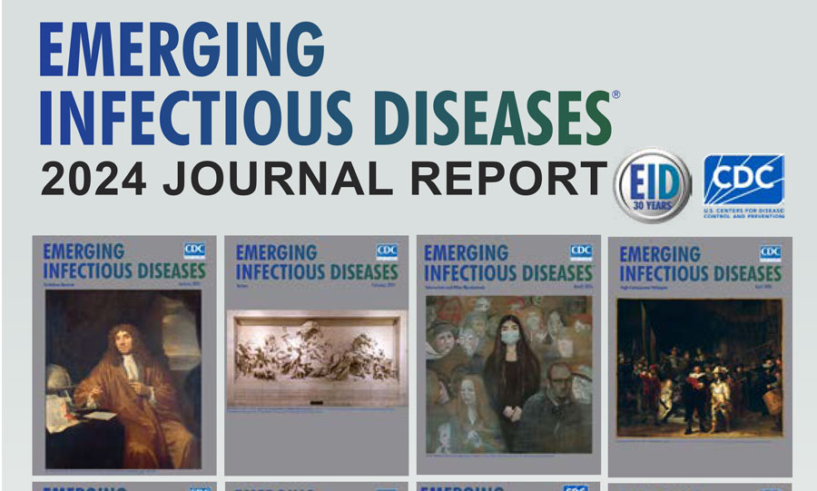
Articles from Emerging Infectious Diseases

Volume 32, Number 12—December 2026
 Podcasts
Synopses
Case Report and Systematic Review of Human Breast Dirofilariasis, 1959–2025 [PDF - 1.40 MB - 10 pages]
Human dirofilariasis is a mosquitoborne zoonosis predominantly caused by Dirofilaria repens nematodes in the Old World; breast involvement is uncommon and mimics breast neoplasia. We report a previously unpublished case from Israel. We also conducted a systematic review of histologically confirmed breast dirofilariasis cases retrieved from PubMed, Embase, and Scopus databases and from searching other literature. In total, we analyzed 40 breast dirofilariasis cases reported during 1959–2025. Most (95%, 38/40) patients were female; all were adults. Most cases were acquired in the Mediterranean rim (53%, 21/40) and India (15%, 6/40). Species-specific diagnosis was commonly made on the basis of morphology alone (37/40); those cases should be considered probable. The most common species was D. repens (n = 31); only 2 cases were molecularly confirmed. Excision was curative in all cases. Breast dirofilariasis should be considered in the differential diagnosis of solitary breast nodules in persons who live in or travel to endemic regions.
| EID | Grupel D, Tenenbaum S, Schwartz E. Case Report and Systematic Review of Human Breast Dirofilariasis, 1959–2025. Emerg Infect Dis. 2026;32(12):1945-1954. https://doi.org/10.3201/eid3212.260994 |
|---|---|
| AMA | Grupel D, Tenenbaum S, Schwartz E. Case Report and Systematic Review of Human Breast Dirofilariasis, 1959–2025. Emerging Infectious Diseases. 2026;32(12):1945-1954. doi:10.3201/eid3212.260994. |
| APA | Grupel, D., Tenenbaum, S., & Schwartz, E. (2026). Case Report and Systematic Review of Human Breast Dirofilariasis, 1959–2025. Emerging Infectious Diseases, 32(12), 1945-1954. https://doi.org/10.3201/eid3212.260994. |
Cryptic Invasion and Transmission Dynamics of Peste des Petits Ruminants Virus, Europe, 2024–2026 [PDF - 3.45 MB - 11 pages]
Peste des petits ruminants (PPR) disease has spread in southeastern Europe since July 2024. Virus sequence analyses and field investigations covering 7 countries affected through January 2026 suggest that eastern Romania was the point of introduction and source of regional viral dissemination in the region. Cross-border transmission inferred from genetic data was strongly associated with official records of live animal trade; outbreak investigations indicate additional roles for illegal trade and indirect transmission. Important temporal gaps in the phylogeny suggest that PPR virus has been circulating undetected in multiple areas in southeastern Europe, as noted by the high variability of clinical signs in infected animals. Those findings highlight a real risk for continued spread across Europe. Increased awareness of the difficulty in detecting the disease, coordinated cross-border surveillance, timely data sharing, and integrated control measures are needed to stop the spread of PPR into Europe.
| EID | Guinat C, Guendouz S, Kwiatek O, Connilhere E, Exbrayat A, Kirtzalidou A, et al. Cryptic Invasion and Transmission Dynamics of Peste des Petits Ruminants Virus, Europe, 2024–2026. Emerg Infect Dis. 2026;32(12):1955-1965. https://doi.org/10.3201/eid3212.261012 |
|---|---|
| AMA | Guinat C, Guendouz S, Kwiatek O, et al. Cryptic Invasion and Transmission Dynamics of Peste des Petits Ruminants Virus, Europe, 2024–2026. Emerging Infectious Diseases. 2026;32(12):1955-1965. doi:10.3201/eid3212.261012. |
| APA | Guinat, C., Guendouz, S., Kwiatek, O., Connilhere, E., Exbrayat, A., Kirtzalidou, A....Bataille, A. (2026). Cryptic Invasion and Transmission Dynamics of Peste des Petits Ruminants Virus, Europe, 2024–2026. Emerging Infectious Diseases, 32(12), 1955-1965. https://doi.org/10.3201/eid3212.261012. |
Research
Identification of Zoonotic Pteropine orthoreovirus in Rousettus amplexicaudatus Fruit Bats, Indonesia [PDF - 18.32 MB - 13 pages]
Pteropine orthoreovirus (PRV) is an emerging batborne virus with zoonotic potential increasingly linked to human respiratory illness. During 2020–2021, we sampled 182 fruit bats from animal markets and suppliers across Java, Indonesia, and identified species morphologically. We inoculated rectal swab samples onto Vero cells, and used next-generation technology to sequence cultures exhibiting cytopathic effects. We recovered 2 isolates from Rousettus amplexicaudatus bats sold for human consumption in Cianjur, West Java. Comparative analysis of 9 of 10 genomic segments showed the isolates were nearly identical (>99%–100%), except for the S2 segment (88.9%). Phylogenetic analysis indicated close clustering with Nelson Bay, Melaka, Kampar, Pulau, and Garut-69 bat-associated PRVs. We found PRV in R. amplexicaudatus bats in Indonesia, which highlights the zoonotic risk of wildlife trade. Continued surveillance of PRVs at the human–bat interface will be needed to assess the emergence of novel viruses and mitigate potential zoonotic threats.
| EID | Dharmayanti N, Nurjanah D, Maladan Y. Identification of Zoonotic Pteropine orthoreovirus in Rousettus amplexicaudatus Fruit Bats, Indonesia. Emerg Infect Dis. 2026;32(12):1966-1978. https://doi.org/10.3201/eid3212.251991 |
|---|---|
| AMA | Dharmayanti N, Nurjanah D, Maladan Y. Identification of Zoonotic Pteropine orthoreovirus in Rousettus amplexicaudatus Fruit Bats, Indonesia. Emerging Infectious Diseases. 2026;32(12):1966-1978. doi:10.3201/eid3212.251991. |
| APA | Dharmayanti, N., Nurjanah, D., & Maladan, Y. (2026). Identification of Zoonotic Pteropine orthoreovirus in Rousettus amplexicaudatus Fruit Bats, Indonesia. Emerging Infectious Diseases, 32(12), 1966-1978. https://doi.org/10.3201/eid3212.251991. |
Estimated Illness Linked to Animal Contact for 7 Pathogens, United States, 2019 [PDF - 555 KB - 9 pages]
Diseases transmitted through direct or indirect animal contact are a public health concern in the United States. Improvements in laboratory detection methods and illness estimates warrant an update of illness estimates linked to animal contact. We used surveillance data and other sources to estimate the annual number of US illnesses, hospitalizations, and deaths for 7 pathogens. We adjusted for underreporting and underdiagnosis by using established methods. Campylobacter, Cryptosporidium, Shiga toxin–producing Escherichia coli, Listeria monocytogenes, nontyphoidal Salmonella, and non-pestis Yersinia caused an estimated 995,022 (90% credible interval [CrI] 476,251–1,833,238) animal contact illnesses around 2019. Together with Toxoplasma gondii, those illnesses caused an estimated 9,592 (90% CrI 5,603‒15,557) hospitalizations and 236 (90% CrI 119‒411) deaths in the United States. Increased awareness among health professionals, government officials, industry partners, and the public about animal contact as a mode of disease transmission can aid prevention efforts and enhance surveillance.
| EID | Medford R, Cui Z, Tierney R, Straily A, Miko S, Francois-Watkins L, et al. Estimated Illness Linked to Animal Contact for 7 Pathogens, United States, 2019. Emerg Infect Dis. 2026;32(12):1979-1987. https://doi.org/10.3201/eid3212.260787 |
|---|---|
| AMA | Medford R, Cui Z, Tierney R, et al. Estimated Illness Linked to Animal Contact for 7 Pathogens, United States, 2019. Emerging Infectious Diseases. 2026;32(12):1979-1987. doi:10.3201/eid3212.260787. |
| APA | Medford, R., Cui, Z., Tierney, R., Straily, A., Miko, S., Francois-Watkins, L....Benedict, K. M. (2026). Estimated Illness Linked to Animal Contact for 7 Pathogens, United States, 2019. Emerging Infectious Diseases, 32(12), 1979-1987. https://doi.org/10.3201/eid3212.260787. |
Detection of Jamestown Canyon Virus in Mosquito Populations, Northeastern Illinois, USA, 2024 [PDF - 1.74 MB - 8 pages]
Jamestown Canyon virus (JCV) is an underreported mosquitoborne arbovirus that can cause viral encephalitis in humans. To determine whether humans in greater Chicago, Illinois, USA, were at risk for JCV infection, we conducted surveillance on mosquito populations in suburban forest preserves with seasonal wetland habitats likely to sustain enzootic transmission. We documented the diversity and abundance of putative vector mosquito species to determine their prevalence for JCV infection. Of 11 predominantly mammalophilic mosquito taxa screened, we detected JCV in 5 species. The highest JCV prevalence was in Anopheles punctipennis, followed by Aedes stimulans group, Ae. canadensis, Ae. trivittatus, and Coquillettidia perturbans mosquitoes. Most detections were from mosquitoes collected in June and early July, and earliest detections were limited to Ae. stimulans group female mosquitoes. Our findings indicate substantial potential for human JCV exposure where urban development encroaches on natural habitats in northeastern Illinois. We urge continued surveillance for JCV.
| EID | Mackay AJ, Cara C, Blackshear M, Jones LE, Jude N, Seo D, et al. Detection of Jamestown Canyon Virus in Mosquito Populations, Northeastern Illinois, USA, 2024. Emerg Infect Dis. 2026;32(12):1988-1995. https://doi.org/10.3201/eid3212.260243 |
|---|---|
| AMA | Mackay AJ, Cara C, Blackshear M, et al. Detection of Jamestown Canyon Virus in Mosquito Populations, Northeastern Illinois, USA, 2024. Emerging Infectious Diseases. 2026;32(12):1988-1995. doi:10.3201/eid3212.260243. |
| APA | Mackay, A. J., Cara, C., Blackshear, M., Jones, L. E., Jude, N., Seo, D....Stone, C. (2026). Detection of Jamestown Canyon Virus in Mosquito Populations, Northeastern Illinois, USA, 2024. Emerging Infectious Diseases, 32(12), 1988-1995. https://doi.org/10.3201/eid3212.260243. |
Monkeypox Virus Seroprevalence among Adults Assessed at Roadside Wellness Centers along Major Transportation Corridor, Kenya, 2025 [PDF - 895 KB - 11 pages]
Since 2024, Kenya has confirmed >1,000 cases of monkeypox virus (MPXV) infection. We conducted a cross-sectional serosurvey at 8 clinics along a transportation corridor in Kenya to quantify unrecognized MPXV transmission. From September 29–October 28, 2025, adults >18 years completed a questionnaire and provided blood samples tested with a serologic assay that distinguishes likely MPXV infection from orthopoxvirus vaccination. We used a modified Poisson regression to assess associations with likely MPXV infection. We compared participants with serologic evidence of MPXV infection to those self-reporting prior confirmed mpox cases. Of 2,896 participants, 332 (11.5%) were seropositive, including 82 (2.8%) classified as likely MPXV infection. Four participants reported prior confirmed MPXV cases, yielding a 21:1 ratio of participants with serologic evidence of MPXV infection to self-reported confirmed cases. Despite some evidence of undetected transmission, most participants lacked serologic evidence of prior orthopoxvirus exposure, indicating a large susceptible population and supporting additional vaccination and ongoing serosurveillance.
| EID | Hines JZ, Lagat D, Kikwai G, Kagecha C, Mwazighe H, Ochieng C, et al. Monkeypox Virus Seroprevalence among Adults Assessed at Roadside Wellness Centers along Major Transportation Corridor, Kenya, 2025. Emerg Infect Dis. 2026;32(12):1996-2006. https://doi.org/10.3201/eid3212.260957 |
|---|---|
| AMA | Hines JZ, Lagat D, Kikwai G, et al. Monkeypox Virus Seroprevalence among Adults Assessed at Roadside Wellness Centers along Major Transportation Corridor, Kenya, 2025. Emerging Infectious Diseases. 2026;32(12):1996-2006. doi:10.3201/eid3212.260957. |
| APA | Hines, J. Z., Lagat, D., Kikwai, G., Kagecha, C., Mwazighe, H., Ochieng, C....Lucchi, N. (2026). Monkeypox Virus Seroprevalence among Adults Assessed at Roadside Wellness Centers along Major Transportation Corridor, Kenya, 2025. Emerging Infectious Diseases, 32(12), 1996-2006. https://doi.org/10.3201/eid3212.260957. |
Rat Hepatitis E Virus Circulation in Brown Rats from 8 Cities, France, 2021–2023 [PDF - 3.62 MB - 12 pages]
Rat hepatitis E virus (rHEV) is a zoonotic pathogen responsible for acute or chronic hepatitis in humans. To assess rHEV circulation in brown rats (Rattus norvegicus) in France, we sampled animals in 8 cities. We detected rHEV RNA in livers of 209/686 rats, a 30.5% positivity rate (95% CI 27.1%–34.0%). We found rHEV-positive animals in all 8 cities; positivity rate range was 9.7% (95% CI 6.7%–13.8%) to 44% (95% CI 33.9%–55.3%). Positivity rate differed by weight class: 9% (95% CI 5.8%–13.8%) in rats <100 g and 40.7% (95% CI 36.3%–45.2%) in rats >100 g. We detected rHEV antibodies in 144/317 (45% [95% CI 40.0%–51.0%]) of the samples. We obtained 20 full genome sequences with distinct rHEV strains. We observed high genetic identity between rat and human sequences in France and Europe. rHEV appears to be enzootic in rat populations in cities in France.
| EID | Pellerin M, Kwasiborski A, Lattard V, Sauvage V, Pisanu B, Hourdel V, et al. Rat Hepatitis E Virus Circulation in Brown Rats from 8 Cities, France, 2021–2023. Emerg Infect Dis. 2026;32(12):2007-2018. https://doi.org/10.3201/eid3212.260926 |
|---|---|
| AMA | Pellerin M, Kwasiborski A, Lattard V, et al. Rat Hepatitis E Virus Circulation in Brown Rats from 8 Cities, France, 2021–2023. Emerging Infectious Diseases. 2026;32(12):2007-2018. doi:10.3201/eid3212.260926. |
| APA | Pellerin, M., Kwasiborski, A., Lattard, V., Sauvage, V., Pisanu, B., Hourdel, V....Pavio, N. (2026). Rat Hepatitis E Virus Circulation in Brown Rats from 8 Cities, France, 2021–2023. Emerging Infectious Diseases, 32(12), 2007-2018. https://doi.org/10.3201/eid3212.260926. |
Dispatches
Isolation of Orientia tsutsugamushi from Bat-Associated Chiggers, Japan [PDF - 1.24 MB - 4 pages]
We isolated Orientia tsutsugamushi bacteria from bat-associated Whartonia prima chiggers collected from Hipposideros turpis bats on Yonaguni Island, Japan. Serial passage, microscopic examination, growth kinetics, and multilocus sequence analysis confirmed bacterial propagation and identity. Our findings highlight an overlooked bat–chigger interface with potential relevance to broader ecologic surveillance of scrub typhus.
| EID | Qiu Y, Kawai K, Jiménez P, Tamura H, Takahashi M, Ohnuma A, et al. Isolation of Orientia tsutsugamushi from Bat-Associated Chiggers, Japan. Emerg Infect Dis. 2026;32(12):2019-2022. https://doi.org/10.3201/eid3212.261156 |
|---|---|
| AMA | Qiu Y, Kawai K, Jiménez P, et al. Isolation of Orientia tsutsugamushi from Bat-Associated Chiggers, Japan. Emerging Infectious Diseases. 2026;32(12):2019-2022. doi:10.3201/eid3212.261156. |
| APA | Qiu, Y., Kawai, K., Jiménez, P., Tamura, H., Takahashi, M., Ohnuma, A....Nakao, R. (2026). Isolation of Orientia tsutsugamushi from Bat-Associated Chiggers, Japan. Emerging Infectious Diseases, 32(12), 2019-2022. https://doi.org/10.3201/eid3212.261156. |
Infectious African Swine Fever Virus in Spray-Dried Porcine Plasma Used as Feed Ingredient, South Korea, 2026 [PDF - 3.08 MB - 5 pages]
We detected African swine fever virus DNA in feed and spray-dried porcine plasma (SDPP) used as a feed ingredient in South Korea. Pigs inoculated with SDPP developed fatal infection. An isolate phylogenetically clustered with South Korea strains. Our findings highlight the need for strengthened risk management of animal-derived feed ingredients like SDPP.
| EID | Hong S, Kwon J, Kim D, Kang H, Kim Y. Infectious African Swine Fever Virus in Spray-Dried Porcine Plasma Used as Feed Ingredient, South Korea, 2026. Emerg Infect Dis. 2026;32(12):2023-2027. https://doi.org/10.3201/eid3212.261380 |
|---|---|
| AMA | Hong S, Kwon J, Kim D, et al. Infectious African Swine Fever Virus in Spray-Dried Porcine Plasma Used as Feed Ingredient, South Korea, 2026. Emerging Infectious Diseases. 2026;32(12):2023-2027. doi:10.3201/eid3212.261380. |
| APA | Hong, S., Kwon, J., Kim, D., Kang, H., & Kim, Y. (2026). Infectious African Swine Fever Virus in Spray-Dried Porcine Plasma Used as Feed Ingredient, South Korea, 2026. Emerging Infectious Diseases, 32(12), 2023-2027. https://doi.org/10.3201/eid3212.261380. |
Typhoid Fever–Like Manifestation of Scrub Typhus in Patient with Myelodysplastic Syndrome, China, 2025 [PDF - 780 KB - 3 pages]
A 79-year-old man in China sought care for fever and syncope and was diagnosed with myelodysplastic syndrome. Bone marrow showed hemophagocytosis and macrophages resembling typhoid cells, but cultures excluded Salmonella infection. Targeted next-generation sequencing identified Orientia tsutsugamushi, and a concealed popliteal eschar was found. Scrub typhus can produce typhoid-like bone marrow morphology.
| EID | Li Y, Wang W, Gao X, Gu Y. Typhoid Fever–Like Manifestation of Scrub Typhus in Patient with Myelodysplastic Syndrome, China, 2025. Emerg Infect Dis. 2026;32(12):2028-2030. https://doi.org/10.3201/eid3212.260686 |
|---|---|
| AMA | Li Y, Wang W, Gao X, et al. Typhoid Fever–Like Manifestation of Scrub Typhus in Patient with Myelodysplastic Syndrome, China, 2025. Emerging Infectious Diseases. 2026;32(12):2028-2030. doi:10.3201/eid3212.260686. |
| APA | Li, Y., Wang, W., Gao, X., & Gu, Y. (2026). Typhoid Fever–Like Manifestation of Scrub Typhus in Patient with Myelodysplastic Syndrome, China, 2025. Emerging Infectious Diseases, 32(12), 2028-2030. https://doi.org/10.3201/eid3212.260686. |
Emergence of Shamonda Orthobunyavirus, Central Europe, 2026 [PDF - 1.59 MB - 5 pages]
Since mid-June 2026, an acute, nonfatal syndrome characterized by high herd illness rate, reduced milk yield, diarrhea, fever, and lethargy has affected dairy cattle in southern Germany and Switzerland. After major viral pathogens were excluded, pan-Simbuvirus reverse transcription PCR detected an orthobunyavirus, confirmed by metagenomic sequencing, enabling recovery of a complete Shamonda virus genome.
| EID | Wernike K, Hoffmann B, Link EK, Rotheneder S, Eshak M, Pfaff F, et al. Emergence of Shamonda Orthobunyavirus, Central Europe, 2026. Emerg Infect Dis. 2026;32(12):2031-2035. https://doi.org/10.3201/eid3212.261496 |
|---|---|
| AMA | Wernike K, Hoffmann B, Link EK, et al. Emergence of Shamonda Orthobunyavirus, Central Europe, 2026. Emerging Infectious Diseases. 2026;32(12):2031-2035. doi:10.3201/eid3212.261496. |
| APA | Wernike, K., Hoffmann, B., Link, E. K., Rotheneder, S., Eshak, M., Pfaff, F....Beer, M. (2026). Emergence of Shamonda Orthobunyavirus, Central Europe, 2026. Emerging Infectious Diseases, 32(12), 2031-2035. https://doi.org/10.3201/eid3212.261496. |
Oropouche Fever Outbreak, Havana, Cuba, June–September 2024 [PDF - 331 KB - 4 pages]
Oropouche fever emerged nationwide in Cuba in 2024. We investigated the outbreak in Havana; of 549 suspected cases, 189 (34.4%) were confirmed. No cases were severe. Incidence was 8.9/100,000 population and was highest in adults 27–59 years of age (10.2/100,000 population). Three infants tested positive. Dengue cocirculation challenged surveillance.
| EID | Martínez-Rodríguez A, Rodríguez E, Díaz A, Vera M, Águila N, Rodríguez W, et al. Oropouche Fever Outbreak, Havana, Cuba, June–September 2024. Emerg Infect Dis. 2026;32(12):2036-2039. https://doi.org/10.3201/eid3212.251334 |
|---|---|
| AMA | Martínez-Rodríguez A, Rodríguez E, Díaz A, et al. Oropouche Fever Outbreak, Havana, Cuba, June–September 2024. Emerging Infectious Diseases. 2026;32(12):2036-2039. doi:10.3201/eid3212.251334. |
| APA | Martínez-Rodríguez, A., Rodríguez, E., Díaz, A., Vera, M., Águila, N., Rodríguez, W....Guzmán, M. G. (2026). Oropouche Fever Outbreak, Havana, Cuba, June–September 2024. Emerging Infectious Diseases, 32(12), 2036-2039. https://doi.org/10.3201/eid3212.251334. |
Wild Mice and Shrews as Natural Reservoirs of Emerging Tickborne Yezo Virus, Hokkaido, Japan, 2023–2025 [PDF - 2.46 MB - 6 pages]
We screened 420 wild small mammals and 2,273 ticks for infections with an emerging tickborne orthonairovirus, Yezo virus, during 2023–2025 in Hokkaido, Japan. We detected virus in mice, shrews, and ticks that were closely related. Apodemus spp. mice and Sorex spp. shrews were identified as potent natural reservoirs of Yezo virus.
| EID | Ito M, Ohari Y, Kojima I, Mimura Y, Mizuma K, Li J, et al. Wild Mice and Shrews as Natural Reservoirs of Emerging Tickborne Yezo Virus, Hokkaido, Japan, 2023–2025. Emerg Infect Dis. 2026;32(12):2040-2045. https://doi.org/10.3201/eid3212.260298 |
|---|---|
| AMA | Ito M, Ohari Y, Kojima I, et al. Wild Mice and Shrews as Natural Reservoirs of Emerging Tickborne Yezo Virus, Hokkaido, Japan, 2023–2025. Emerging Infectious Diseases. 2026;32(12):2040-2045. doi:10.3201/eid3212.260298. |
| APA | Ito, M., Ohari, Y., Kojima, I., Mimura, Y., Mizuma, K., Li, J....Matsuno, K. (2026). Wild Mice and Shrews as Natural Reservoirs of Emerging Tickborne Yezo Virus, Hokkaido, Japan, 2023–2025. Emerging Infectious Diseases, 32(12), 2040-2045. https://doi.org/10.3201/eid3212.260298. |
Mayaro Virus Emergence and Genetic Dynamics, Peru [PDF - 1.53 MB - 5 pages]
Mayaro virus (MAYV) is an emerging pathogen in Latin America. Using phylodynamic reconstructions and phylowave analyses, we describe novel lineage diversification and rapid dispersal of MAYV in Peru. A newly developed, dual-target quantitative reverse transcription PCR assay reliably detects MAYV variants, enabling improved surveillance, control, and understanding of this neglected arbovirus.
| EID | Perez LJ, Hodges A, Torres K, Forberg K, Weiss S, Yamaguchi J, et al. Mayaro Virus Emergence and Genetic Dynamics, Peru. Emerg Infect Dis. 2026;32(12):2046-2050. https://doi.org/10.3201/eid3212.251211 |
|---|---|
| AMA | Perez LJ, Hodges A, Torres K, et al. Mayaro Virus Emergence and Genetic Dynamics, Peru. Emerging Infectious Diseases. 2026;32(12):2046-2050. doi:10.3201/eid3212.251211. |
| APA | Perez, L. J., Hodges, A., Torres, K., Forberg, K., Weiss, S., Yamaguchi, J....Cloherty, G. A. (2026). Mayaro Virus Emergence and Genetic Dynamics, Peru. Emerging Infectious Diseases, 32(12), 2046-2050. https://doi.org/10.3201/eid3212.251211. |
Natural Occurrence of Primate SLAM-Adapted Canine Distemper Virus, Brazil, 2018 [PDF - 4.30 MB - 6 pages]
We analyzed canine distemper virus (CDV) from a 2018 marmoset disease outbreak in Brazil. CDV hemagglutinin protein bound to the marmoset SLAM (signaling lymphocytic activation molecule) receptor through 2 amino acid substitutions within hemagglutinin, which also enhanced human SLAM receptor usage. Continued surveillance will be critical to monitor CDV cross-species transmission.
| EID | Tominaga M, Hyodo A, Kojima S, Onodera M, Yang H, Seki F, et al. Natural Occurrence of Primate SLAM-Adapted Canine Distemper Virus, Brazil, 2018. Emerg Infect Dis. 2026;32(12):2051-2056. https://doi.org/10.3201/eid3212.260946 |
|---|---|
| AMA | Tominaga M, Hyodo A, Kojima S, et al. Natural Occurrence of Primate SLAM-Adapted Canine Distemper Virus, Brazil, 2018. Emerging Infectious Diseases. 2026;32(12):2051-2056. doi:10.3201/eid3212.260946. |
| APA | Tominaga, M., Hyodo, A., Kojima, S., Onodera, M., Yang, H., Seki, F....Takeda, M. (2026). Natural Occurrence of Primate SLAM-Adapted Canine Distemper Virus, Brazil, 2018. Emerging Infectious Diseases, 32(12), 2051-2056. https://doi.org/10.3201/eid3212.260946. |
Molecular Surveillance for Oropouche, Mayaro, West Nile, and Saint Louis EncephalitisViruses, Brazil, 2019–2025 [PDF - 1.01 MB - 5 pages]
To assess the circulation of Oropouche, Mayaro, West Nile, and St. Louis encephalitis viruses, we tested samples from 824 patients with acute febrile illness, 3,484 mosquitoes, and 113 nonhuman primates from northern Minas Gerais, Brazil. All samples were negative for dengue, Chikungunya, and Zika virus. One imported Oropouche virus infection was detected.
| EID | Ferreira A, Teixeira TJ, de Oliveira DB, Schetino M, de Oliveira CH, Souza P, et al. Molecular Surveillance for Oropouche, Mayaro, West Nile, and Saint Louis EncephalitisViruses, Brazil, 2019–2025. Emerg Infect Dis. 2026;32(12):2057-2061. https://doi.org/10.3201/eid3212.260870 |
|---|---|
| AMA | Ferreira A, Teixeira TJ, de Oliveira DB, et al. Molecular Surveillance for Oropouche, Mayaro, West Nile, and Saint Louis EncephalitisViruses, Brazil, 2019–2025. Emerging Infectious Diseases. 2026;32(12):2057-2061. doi:10.3201/eid3212.260870. |
| APA | Ferreira, A., Teixeira, T. J., de Oliveira, D. B., Schetino, M., de Oliveira, C. H., Souza, P....Rocha-Vieira, E. (2026). Molecular Surveillance for Oropouche, Mayaro, West Nile, and Saint Louis EncephalitisViruses, Brazil, 2019–2025. Emerging Infectious Diseases, 32(12), 2057-2061. https://doi.org/10.3201/eid3212.260870. |
Leptospira Exposure in Urban River Surfers, Germany, 2024 [PDF - 1.04 MB - 5 pages]
After 4 urban river surfers in Munich, Germany, were hospitalized with severe leptospirosis, we conducted a controlled cross-sectional study. Leptospira IgG seroreactivity was more frequent among surfers than controls, and Leptospira DNA was detected in 1 soil sample. Freshwater surfing could represent an underrecognized exposure setting in temperate cities.
| EID | Voit F, Schult-Hannemann D, Wagner L, Ostermaier J, Stollberg K, Luge E, et al. Leptospira Exposure in Urban River Surfers, Germany, 2024. Emerg Infect Dis. 2026;32(12):2062-2066. https://doi.org/10.3201/eid3212.260879 |
|---|---|
| AMA | Voit F, Schult-Hannemann D, Wagner L, et al. Leptospira Exposure in Urban River Surfers, Germany, 2024. Emerging Infectious Diseases. 2026;32(12):2062-2066. doi:10.3201/eid3212.260879. |
| APA | Voit, F., Schult-Hannemann, D., Wagner, L., Ostermaier, J., Stollberg, K., Luge, E....Richter, M. H. (2026). Leptospira Exposure in Urban River Surfers, Germany, 2024. Emerging Infectious Diseases, 32(12), 2062-2066. https://doi.org/10.3201/eid3212.260879. |
Post–COVID-19 Emergence and Spread of Hypertransmissible Cryptosporidium hominis gp60 Subtype IfA12G1R5, Spain [PDF - 899 KB - 5 pages]
COVID-19 pandemic containment measures in 2020 selectively altered established Cryptosporidium hominis anthroponotic transmission in Spain. Rapid emergence and expansion of hypertransmissible human-adapted gp60 subtypes such as IfA12G1R5 and, less markedly, IbA13G3 followed. Conversely, transmission of the largely zoonotic C. parvum remained comparatively stable, yielding a dual epidemiologic scenario in this country.
| EID | Goterris L, Köster PC, Fuentes J, Sulleiro E, Alguacil-Guillén M, Álvarez-Martínez MJ, et al. Post–COVID-19 Emergence and Spread of Hypertransmissible Cryptosporidium hominis gp60 Subtype IfA12G1R5, Spain. Emerg Infect Dis. 2026;32(12):2067-2071. https://doi.org/10.3201/eid3212.261007 |
|---|---|
| AMA | Goterris L, Köster PC, Fuentes J, et al. Post–COVID-19 Emergence and Spread of Hypertransmissible Cryptosporidium hominis gp60 Subtype IfA12G1R5, Spain. Emerging Infectious Diseases. 2026;32(12):2067-2071. doi:10.3201/eid3212.261007. |
| APA | Goterris, L., Köster, P. C., Fuentes, J., Sulleiro, E., Alguacil-Guillén, M., Álvarez-Martínez, M. J....Carmena, D. (2026). Post–COVID-19 Emergence and Spread of Hypertransmissible Cryptosporidium hominis gp60 Subtype IfA12G1R5, Spain. Emerging Infectious Diseases, 32(12), 2067-2071. https://doi.org/10.3201/eid3212.261007. |
Endemic Cryptococcus gattii Infections, Nova Scotia, Canada, 2016–2024 [PDF - 353 KB - 3 pages]
Cryptococcus gattii is poorly characterized in Atlantic Canada. We identified 6 species-confirmed infections in Nova Scotia during 2016–2024. Of those, 5 in western Nova Scotia were genotype VGIIb, including 4 cases without travel outside the province. Previous VGIIb-like detection in a wild animal further supports local acquisition and regional endemicity.
| EID | Zayed R, DiQuinzio M, Patriquin G, Simms E, O’Brien K, Robinson A, et al. Endemic Cryptococcus gattii Infections, Nova Scotia, Canada, 2016–2024. Emerg Infect Dis. 2026;32(12):2072-2074. https://doi.org/10.3201/eid3212.260971 |
|---|---|
| AMA | Zayed R, DiQuinzio M, Patriquin G, et al. Endemic Cryptococcus gattii Infections, Nova Scotia, Canada, 2016–2024. Emerging Infectious Diseases. 2026;32(12):2072-2074. doi:10.3201/eid3212.260971. |
| APA | Zayed, R., DiQuinzio, M., Patriquin, G., Simms, E., O’Brien, K., Robinson, A....McIsaac, K. (2026). Endemic Cryptococcus gattii Infections, Nova Scotia, Canada, 2016–2024. Emerging Infectious Diseases, 32(12), 2072-2074. https://doi.org/10.3201/eid3212.260971. |
Livestock-Associated Methicillin-Susceptible Staphylococcus aureus Bacteremia, Denmark, 2007–2023 [PDF - 649 KB - 5 pages]
Livestock-associated Staphylococcus aureus caused 200 human bloodstream infections in Denmark during 2007–2023; two thirds were caused by methicillin-susceptible isolates. Risk for death was comparable to that associated with human S. aureus clones. Industrial pig population size correlated strongly with disease prevalence. Seventeen percent of isolates showed signs of human adaptation.
| EID | Raisen CL, Pedersen AB, Dalsgaard A, Mølbak K, Petersen A, Larsen AR, et al. Livestock-Associated Methicillin-Susceptible Staphylococcus aureus Bacteremia, Denmark, 2007–2023. Emerg Infect Dis. 2026;32(12):2075-2079. https://doi.org/10.3201/eid3212.261261 |
|---|---|
| AMA | Raisen CL, Pedersen AB, Dalsgaard A, et al. Livestock-Associated Methicillin-Susceptible Staphylococcus aureus Bacteremia, Denmark, 2007–2023. Emerging Infectious Diseases. 2026;32(12):2075-2079. doi:10.3201/eid3212.261261. |
| APA | Raisen, C. L., Pedersen, A. B., Dalsgaard, A., Mølbak, K., Petersen, A., Larsen, A. R....Larsen, J. (2026). Livestock-Associated Methicillin-Susceptible Staphylococcus aureus Bacteremia, Denmark, 2007–2023. Emerging Infectious Diseases, 32(12), 2075-2079. https://doi.org/10.3201/eid3212.261261. |
Indirect Healthcare-Associated Canine Sporotrichosis Caused by Sporothrix brasiliensis, Argentina, 2024 [PDF - 1.50 MB - 6 pages]
We describe a Sporothrix brasiliensis infection in a dog in Puerto Iguazú, Argentina, in 2024, that clustered with 3 epidemiologically linked feline sporotrichosis cases at the same veterinary clinic. Early mycologic diagnosis, molecular surveillance, and rigorous infection-control practices are critical to prevent healthcare-associated S. brasiliensis exposures in veterinary settings.
| EID | Vizcaychipi KA, López-Joffre MC, Couto EM, Gremião I, Mendoza JA, Viale MN, et al. Indirect Healthcare-Associated Canine Sporotrichosis Caused by Sporothrix brasiliensis, Argentina, 2024. Emerg Infect Dis. 2026;32(12):2080-2085. https://doi.org/10.3201/eid3212.261410 |
|---|---|
| AMA | Vizcaychipi KA, López-Joffre MC, Couto EM, et al. Indirect Healthcare-Associated Canine Sporotrichosis Caused by Sporothrix brasiliensis, Argentina, 2024. Emerging Infectious Diseases. 2026;32(12):2080-2085. doi:10.3201/eid3212.261410. |
| APA | Vizcaychipi, K. A., López-Joffre, M. C., Couto, E. M., Gremião, I., Mendoza, J. A., Viale, M. N....Canteros, C. E. (2026). Indirect Healthcare-Associated Canine Sporotrichosis Caused by Sporothrix brasiliensis, Argentina, 2024. Emerging Infectious Diseases, 32(12), 2080-2085. https://doi.org/10.3201/eid3212.261410. |
Rat Hepatitis E Virus and Seoul Hantavirus in Pet Rats, Germany [PDF - 1.30 MB - 4 pages]
Pet rats represent a reservoir of multiple zoonotic pathogens. We demonstrated that pet rats, previously confirmed to be Seoul orthohantavirus–infected and involved in human infections in Germany, were concurrently infected with rat hepatitis E virus. The high degree of rat hepatitis E virus sequence identity suggests a single incursion event.
| EID | Mehl C, Schilling-Loeffler K, Haake A, Sehl-Ewert J, Schmidt K, de Cock MP, et al. Rat Hepatitis E Virus and Seoul Hantavirus in Pet Rats, Germany. Emerg Infect Dis. 2026;32(12):2086-2089. https://doi.org/10.3201/eid3212.261208 |
|---|---|
| AMA | Mehl C, Schilling-Loeffler K, Haake A, et al. Rat Hepatitis E Virus and Seoul Hantavirus in Pet Rats, Germany. Emerging Infectious Diseases. 2026;32(12):2086-2089. doi:10.3201/eid3212.261208. |
| APA | Mehl, C., Schilling-Loeffler, K., Haake, A., Sehl-Ewert, J., Schmidt, K., de Cock, M. P....Ulrich, R. G. (2026). Rat Hepatitis E Virus and Seoul Hantavirus in Pet Rats, Germany. Emerging Infectious Diseases, 32(12), 2086-2089. https://doi.org/10.3201/eid3212.261208. |
Detection of Mycobacterium africanum Lineage 6 in White-Nosed Coatis, Costa Rica [PDF - 2.08 MB - 5 pages]
We detected disseminated granulomatous disease with abundant acid-fast bacilli in free-ranging, white-nosed coatis in Costa Rica. Molecular characterization identified Mycobacterium africanum lineage 6. Our findings demonstrate the presence of M. africanum in free-ranging wildlife in the Americas and highlight the value of wildlife surveillance for prompting coordinated One Health investigation and response.
| EID | Aguilar-Vargas F, Solorzano-Scott T, Hutter SE, Cordero-Salas M, Gazel N, Espinoza S, et al. Detection of Mycobacterium africanum Lineage 6 in White-Nosed Coatis, Costa Rica. Emerg Infect Dis. 2026;32(12):2090-2094. https://doi.org/10.3201/eid3212.260362 |
|---|---|
| AMA | Aguilar-Vargas F, Solorzano-Scott T, Hutter SE, et al. Detection of Mycobacterium africanum Lineage 6 in White-Nosed Coatis, Costa Rica. Emerging Infectious Diseases. 2026;32(12):2090-2094. doi:10.3201/eid3212.260362. |
| APA | Aguilar-Vargas, F., Solorzano-Scott, T., Hutter, S. E., Cordero-Salas, M., Gazel, N., Espinoza, S....Baldi, M. (2026). Detection of Mycobacterium africanum Lineage 6 in White-Nosed Coatis, Costa Rica. Emerging Infectious Diseases, 32(12), 2090-2094. https://doi.org/10.3201/eid3212.260362. |
Research Letters
Zoonotic Exposure to Porcine Deltacoronavirus, United States, 2019–2021 [PDF - 795 KB - 3 pages]
Porcine deltacoronavirus (PDCoV) is a swine coronavirus with zoonotic potential. We detected PDCoV-neutralizing antibodies in 10 of 300 human serum samples; positive sample sources included 1 adult and 9 juveniles. Our findings provide evidence of human exposure to PDCoV and highlight the importance of monitoring animal coronaviruses with zoonotic potential.
| EID | Boley PA, Hartley G, Saif LJ, Bowman AS, Nolting JM, Kenney SP. Zoonotic Exposure to Porcine Deltacoronavirus, United States, 2019–2021. Emerg Infect Dis. 2026;32(12):2107-2109. https://doi.org/10.3201/eid3212.260303 |
|---|---|
| AMA | Boley PA, Hartley G, Saif LJ, et al. Zoonotic Exposure to Porcine Deltacoronavirus, United States, 2019–2021. Emerging Infectious Diseases. 2026;32(12):2107-2109. doi:10.3201/eid3212.260303. |
| APA | Boley, P. A., Hartley, G., Saif, L. J., Bowman, A. S., Nolting, J. M., & Kenney, S. P. (2026). Zoonotic Exposure to Porcine Deltacoronavirus, United States, 2019–2021. Emerging Infectious Diseases, 32(12), 2107-2109. https://doi.org/10.3201/eid3212.260303. |
Fatal Disseminated Powassan Virus Infection in Transplant Recipient, Pennsylvania, USA [PDF - 1.35 MB - 4 pages]
We describe histopathologic findings from a fatal case of Powassan virus lineage 2 (deer tick virus) in an immunosuppressed liver transplant patient in Pennsylvania, USA. We confirmed infection in postmortem brain, lung, and kidney by immunohistochemistry, in situ hybridization, and reverse transcription PCR, demonstrating infection of pulmonary and renal tissues.
| EID | Sung DC, Colbourn R, Khan N, DiRienzi J, Randolph L, Valdivieso F, et al. Fatal Disseminated Powassan Virus Infection in Transplant Recipient, Pennsylvania, USA. Emerg Infect Dis. 2026;32(12):2110-2113. https://doi.org/10.3201/eid3212.260193 |
|---|---|
| AMA | Sung DC, Colbourn R, Khan N, et al. Fatal Disseminated Powassan Virus Infection in Transplant Recipient, Pennsylvania, USA. Emerging Infectious Diseases. 2026;32(12):2110-2113. doi:10.3201/eid3212.260193. |
| APA | Sung, D. C., Colbourn, R., Khan, N., DiRienzi, J., Randolph, L., Valdivieso, F....Nasrallah, M. P. (2026). Fatal Disseminated Powassan Virus Infection in Transplant Recipient, Pennsylvania, USA. Emerging Infectious Diseases, 32(12), 2110-2113. https://doi.org/10.3201/eid3212.260193. |
Neoehrlichia mikurensis Infection in Immunocompromised Patient, the Netherlands, 2024 [PDF - 719 KB - 3 pages]
We report a case of Neoehrlichia mikurensis infection from a tick bite in a 69-year-old immunocompromised man with non-Hodgkin lymphoma in the Netherlands. Because of increasing use of immunosuppressive therapies and frequent tick bites, N. mikurensis should be recognized as a potential cause of recurrent fever in immunocompromised patients.
| EID | Tjan M, Boogers WJ, Verweij JJ, Berrevoets M, Hovius JW. Neoehrlichia mikurensis Infection in Immunocompromised Patient, the Netherlands, 2024. Emerg Infect Dis. 2026;32(12):2113-2115. https://doi.org/10.3201/eid3212.261316 |
|---|---|
| AMA | Tjan M, Boogers WJ, Verweij JJ, et al. Neoehrlichia mikurensis Infection in Immunocompromised Patient, the Netherlands, 2024. Emerging Infectious Diseases. 2026;32(12):2113-2115. doi:10.3201/eid3212.261316. |
| APA | Tjan, M., Boogers, W. J., Verweij, J. J., Berrevoets, M., & Hovius, J. W. (2026). Neoehrlichia mikurensis Infection in Immunocompromised Patient, the Netherlands, 2024. Emerging Infectious Diseases, 32(12), 2113-2115. https://doi.org/10.3201/eid3212.261316. |
Serologic Evidence of Avian Influenza Virus Exposure in Livestock, Egypt, 2024–2026 [PDF - 470 KB - 3 pages]
Spillover of influenza A(H5N1) virus to cattle in the United States sparked global concern. We investigated whether endemic avian influenza viruses are potentially spilling over into domestic livestock in Egypt. We detected influenza H9N2 antibodies in 2.5% and H5N1 antibodies in 2.9%–4.5% of tested animals. Additional surveillance will elucidate host infection dynamics.
| EID | Gomaa M, El-Shesheny R, El Taweel A, Kutkat O, Emam H, Helal B, et al. Serologic Evidence of Avian Influenza Virus Exposure in Livestock, Egypt, 2024–2026. Emerg Infect Dis. 2026;32(12):2116-2118. https://doi.org/10.3201/eid3212.260869 |
|---|---|
| AMA | Gomaa M, El-Shesheny R, El Taweel A, et al. Serologic Evidence of Avian Influenza Virus Exposure in Livestock, Egypt, 2024–2026. Emerging Infectious Diseases. 2026;32(12):2116-2118. doi:10.3201/eid3212.260869. |
| APA | Gomaa, M., El-Shesheny, R., El Taweel, A., Kutkat, O., Emam, H., Helal, B....Kayali, G. (2026). Serologic Evidence of Avian Influenza Virus Exposure in Livestock, Egypt, 2024–2026. Emerging Infectious Diseases, 32(12), 2116-2118. https://doi.org/10.3201/eid3212.260869. |
Human Armillifer armillatus Infection Mimicking Strangulated Umbilical Hernia, France, 2026 [PDF - 709 KB - 3 pages]
We report a case of Armillifer armillatus infection in Arras, France, in a woman originally from the Democratic Republic of the Congo. She manifested abdominal pain that mimicked a strangulated umbilical hernia. Clinicians should be aware of this parasitic infection, particularly in patients with a history of snake exposure.
| EID | Devaux J, Fleuriet J, Leroy C, Gangneux J, Delaforge K, Autier B. Human Armillifer armillatus Infection Mimicking Strangulated Umbilical Hernia, France, 2026. Emerg Infect Dis. 2026;32(12):2118-2120. https://doi.org/10.3201/eid3212.261471 |
|---|---|
| AMA | Devaux J, Fleuriet J, Leroy C, et al. Human Armillifer armillatus Infection Mimicking Strangulated Umbilical Hernia, France, 2026. Emerging Infectious Diseases. 2026;32(12):2118-2120. doi:10.3201/eid3212.261471. |
| APA | Devaux, J., Fleuriet, J., Leroy, C., Gangneux, J., Delaforge, K., & Autier, B. (2026). Human Armillifer armillatus Infection Mimicking Strangulated Umbilical Hernia, France, 2026. Emerging Infectious Diseases, 32(12), 2118-2120. https://doi.org/10.3201/eid3212.261471. |
Emerging Protoparvovirus in Pigs and Wildlife, Denmark [PDF - 585 KB - 4 pages]
We detected a protoparvovirus recently associated with an outbreak in pigs in the Netherlands in pigs with exophthalmos and in fox feces and spleens in Denmark. Pig viruses were phylogenetically closer to each other than to viruses found among wildlife of their respective countries, suggesting a link between the farm cases.
| EID | Canuti M, Høeg F, Hammer A, Jensen T, Quaade M, Ryt-Hansen P, et al. Emerging Protoparvovirus in Pigs and Wildlife, Denmark. Emerg Infect Dis. 2026;32(12):2120-2123. https://doi.org/10.3201/eid3212.261184 |
|---|---|
| AMA | Canuti M, Høeg F, Hammer A, et al. Emerging Protoparvovirus in Pigs and Wildlife, Denmark. Emerging Infectious Diseases. 2026;32(12):2120-2123. doi:10.3201/eid3212.261184. |
| APA | Canuti, M., Høeg, F., Hammer, A., Jensen, T., Quaade, M., Ryt-Hansen, P....Larsen, L. (2026). Emerging Protoparvovirus in Pigs and Wildlife, Denmark. Emerging Infectious Diseases, 32(12), 2120-2123. https://doi.org/10.3201/eid3212.261184. |
Heartland Virus in Lone Star Ticks, South Carolina, USA, 2021–2024 [PDF - 584 KB - 4 pages]
Tickborne arboviruses are a serious health threat, but surveillance gaps remain. We tested lone star nymphs collected in South Carolina, USA, for Heartland and Bourbon viruses. Six pools were positive for Heartland virus; none were positive for Bourbon virus. Longitudinal surveillance provides insights into the emergence and distribution of tickborne pathogens.
| EID | Pickle E, Torelli M, Knapp H, Zychowski DL, Sweeney S, Bramlett K, et al. Heartland Virus in Lone Star Ticks, South Carolina, USA, 2021–2024. Emerg Infect Dis. 2026;32(12):2123-2126. https://doi.org/10.3201/eid3212.260320 |
|---|---|
| AMA | Pickle E, Torelli M, Knapp H, et al. Heartland Virus in Lone Star Ticks, South Carolina, USA, 2021–2024. Emerging Infectious Diseases. 2026;32(12):2123-2126. doi:10.3201/eid3212.260320. |
| APA | Pickle, E., Torelli, M., Knapp, H., Zychowski, D. L., Sweeney, S., Bramlett, K....Nolan, M. S. (2026). Heartland Virus in Lone Star Ticks, South Carolina, USA, 2021–2024. Emerging Infectious Diseases, 32(12), 2123-2126. https://doi.org/10.3201/eid3212.260320. |
Osteomyelitis Caused by Metamycoplasma gateae after Cat Bite, Japan, 2026 [PDF - 468 KB - 3 pages]
We report a case of osteomyelitis caused by Metamycoplasma gateae after a cat bite in an immunocompromised patient in Japan. No standardized treatment has been established; however, the patient recovered after surgery and minocycline treatment. Clinicians should consider M. gateae infection in immunocompromised patients with unidentified infections and cat-bite exposure.
| EID | Hashimoto T, Uehara M, Shingai Y, Tamura D, Kimitsuki K, Shibata H, et al. Osteomyelitis Caused by Metamycoplasma gateae after Cat Bite, Japan, 2026. Emerg Infect Dis. 2026;32(12):2126-2128. https://doi.org/10.3201/eid3212.260515 |
|---|---|
| AMA | Hashimoto T, Uehara M, Shingai Y, et al. Osteomyelitis Caused by Metamycoplasma gateae after Cat Bite, Japan, 2026. Emerging Infectious Diseases. 2026;32(12):2126-2128. doi:10.3201/eid3212.260515. |
| APA | Hashimoto, T., Uehara, M., Shingai, Y., Tamura, D., Kimitsuki, K., Shibata, H....Hiramatsu, K. (2026). Osteomyelitis Caused by Metamycoplasma gateae after Cat Bite, Japan, 2026. Emerging Infectious Diseases, 32(12), 2126-2128. https://doi.org/10.3201/eid3212.260515. |
Cross-Protection against Influenza A(H5) Clade 2.3.4.4b Viruses after A(H1N1)pdm09 Infection [PDF - 1.20 MB - 4 pages]
We challenged ferrets previously infected with influenza A(H1N1)pdm09 virus with H5N1 or H5N5 clade 2.3.4.4b viruses to assess cross-protection. Results show that previous A(H1N1)pdm09 virus immunity conferred partial protection against both viruses, with slightly reduced protection against H5N5 compared with H5N1, potentially reflecting the absence of cross-reactive N5 neuraminidase antibodies.
| EID | Sun X, Belser JA, Li Z, Brock N, Pulit-Penaloza JA, Kieran TJ, et al. Cross-Protection against Influenza A(H5) Clade 2.3.4.4b Viruses after A(H1N1)pdm09 Infection. Emerg Infect Dis. 2026;32(12):2128-2131. https://doi.org/10.3201/eid3212.261032 |
|---|---|
| AMA | Sun X, Belser JA, Li Z, et al. Cross-Protection against Influenza A(H5) Clade 2.3.4.4b Viruses after A(H1N1)pdm09 Infection. Emerging Infectious Diseases. 2026;32(12):2128-2131. doi:10.3201/eid3212.261032. |
| APA | Sun, X., Belser, J. A., Li, Z., Brock, N., Pulit-Penaloza, J. A., Kieran, T. J....Maines, T. R. (2026). Cross-Protection against Influenza A(H5) Clade 2.3.4.4b Viruses after A(H1N1)pdm09 Infection. Emerging Infectious Diseases, 32(12), 2128-2131. https://doi.org/10.3201/eid3212.261032. |
Natural Trichuris trichiura Infections in Endemic Hydromyini Rodents, Papua New Guinea, 2019–2023 [PDF - 1.20 MB - 4 pages]
Screening of 874 wild mammals in Papua New Guinea during 2019–2023 identified Trichuris whipworm infections in multiple rodent species. Mitochondrial and nuclear markers identified 8 Trichuris trichiura infections. Those findings broaden the recognized host range of this human soil-transmitted helminth and raise questions regarding wildlife involvement in its transmission ecology.
| EID | Moradpour N, Vejmělka F, Okena D, Novotný V, Bryjová A, Čížková D, et al. Natural Trichuris trichiura Infections in Endemic Hydromyini Rodents, Papua New Guinea, 2019–2023. Emerg Infect Dis. 2026;32(12):2131-2134. https://doi.org/10.3201/eid3212.261101 |
|---|---|
| AMA | Moradpour N, Vejmělka F, Okena D, et al. Natural Trichuris trichiura Infections in Endemic Hydromyini Rodents, Papua New Guinea, 2019–2023. Emerging Infectious Diseases. 2026;32(12):2131-2134. doi:10.3201/eid3212.261101. |
| APA | Moradpour, N., Vejmělka, F., Okena, D., Novotný, V., Bryjová, A., Čížková, D....de Bellocq, J. (2026). Natural Trichuris trichiura Infections in Endemic Hydromyini Rodents, Papua New Guinea, 2019–2023. Emerging Infectious Diseases, 32(12), 2131-2134. https://doi.org/10.3201/eid3212.261101. |
Detection of Mycobacterium ulcerans DNA in African Fruit Bats, Cameroon, 2024 [PDF - 510 KB - 3 pages]
In 2024, we detected Mycobacterium ulcerans DNA in 2 of 46 fruit bats in Akonolinga, Cameroon, whereas 111 aquatic macroinvertebrate pools were negative for M. ulcerans. Broader longitudinal surveillance will be needed to clarify the role of fruit bats as a reservoir for M. ulcerans circulation in Cameroon and Africa.
| EID | Tchatchouang S, Meyin A Ebong S, Baumy C, Robbe-Saule M, Wultof N, Woguia G, et al. Detection of Mycobacterium ulcerans DNA in African Fruit Bats, Cameroon, 2024. Emerg Infect Dis. 2026;32(12):2134-2137. https://doi.org/10.3201/eid3212.261277 |
|---|---|
| AMA | Tchatchouang S, Meyin A Ebong S, Baumy C, et al. Detection of Mycobacterium ulcerans DNA in African Fruit Bats, Cameroon, 2024. Emerging Infectious Diseases. 2026;32(12):2134-2137. doi:10.3201/eid3212.261277. |
| APA | Tchatchouang, S., Meyin A Ebong, S., Baumy, C., Robbe-Saule, M., Wultof, N., Woguia, G....Marsollier, L. (2026). Detection of Mycobacterium ulcerans DNA in African Fruit Bats, Cameroon, 2024. Emerging Infectious Diseases, 32(12), 2134-2137. https://doi.org/10.3201/eid3212.261277. |
Human Discovertebral Infection Caused by Camelimonas Species Bacteria, France, 2025 [PDF - 1.37 MB - 4 pages]
We describe a case of human Camelimonas species infection, highlighting this bacterium as a potential emerging zoonotic pathogen with bone tropism. Diagnosis required repeated biopsies and genome sequencing. The isolate was susceptible to trimethoprim/sulfamethoxazole and doxycycline; clinical improvement followed prolonged therapy. Our findings support this bacterium’s pathogenic role in humans.
| EID | Chombart L, Blosse A, Desclaux A, Jehanne Q, Dauchy F, Neau D, et al. Human Discovertebral Infection Caused by Camelimonas Species Bacteria, France, 2025. Emerg Infect Dis. 2026;32(12):2137-2140. https://doi.org/10.3201/eid3212.260924 |
|---|---|
| AMA | Chombart L, Blosse A, Desclaux A, et al. Human Discovertebral Infection Caused by Camelimonas Species Bacteria, France, 2025. Emerging Infectious Diseases. 2026;32(12):2137-2140. doi:10.3201/eid3212.260924. |
| APA | Chombart, L., Blosse, A., Desclaux, A., Jehanne, Q., Dauchy, F., Neau, D....Brousse, X. (2026). Human Discovertebral Infection Caused by Camelimonas Species Bacteria, France, 2025. Emerging Infectious Diseases, 32(12), 2137-2140. https://doi.org/10.3201/eid3212.260924. |
Letters
New World Ocular Dirofilariasis Caused by Dirofilaria repens Infection, United States [PDF - 267 KB - 1 page]
| EID | Dantas-Torres F, Otranto D. New World Ocular Dirofilariasis Caused by Dirofilaria repens Infection, United States. Emerg Infect Dis. 2026;32(12):2141. https://doi.org/10.3201/eid3212.261388 |
|---|---|
| AMA | Dantas-Torres F, Otranto D. New World Ocular Dirofilariasis Caused by Dirofilaria repens Infection, United States. Emerging Infectious Diseases. 2026;32(12):2141. doi:10.3201/eid3212.261388. |
| APA | Dantas-Torres, F., & Otranto, D. (2026). New World Ocular Dirofilariasis Caused by Dirofilaria repens Infection, United States. Emerging Infectious Diseases, 32(12), 2141. https://doi.org/10.3201/eid3212.261388. |
Another Dimension
An Old Enemy Returns [PDF - 859 KB - 3 pages]
A retired veterinarian reflects on the 2023–2026 reemergence of New World screwworm, from foreign animal disease training exercises decades ago to the 2026 outbreak in South Texas, USA. Correcting common misconceptions about the parasite, he argues that eradication is a maintenance obligation, not a permanent achievement, requiring sustained investment and cooperation.
| EID | Campagnolo ER. An Old Enemy Returns. Emerg Infect Dis. 2026;32(12):2095-2097. https://doi.org/10.3201/eid3212.261043 |
|---|---|
| AMA | Campagnolo ER. An Old Enemy Returns. Emerging Infectious Diseases. 2026;32(12):2095-2097. doi:10.3201/eid3212.261043. |
| APA | Campagnolo, E. R. (2026). An Old Enemy Returns. Emerging Infectious Diseases, 32(12), 2095-2097. https://doi.org/10.3201/eid3212.261043. |
Using Cover Art from Emerging Infectious Diseases to Inspire Microbiological Education [PDF - 2.44 MB - 9 pages]
The Emerging Infectious Diseases journal covers demonstrate how art can depict, explain, and humanize science. Inspired by those works, this article shares teaching strategies to integrate art into microbiological education. With this curriculum, I encourage others to explore how art can deepen our understanding of microbiology and the world around us.
| EID | Iverson-Cabral SL. Using Cover Art from Emerging Infectious Diseases to Inspire Microbiological Education. Emerg Infect Dis. 2026;32(12):2098-2106. https://doi.org/10.3201/eid3212.260532 |
|---|---|
| AMA | Iverson-Cabral SL. Using Cover Art from Emerging Infectious Diseases to Inspire Microbiological Education. Emerging Infectious Diseases. 2026;32(12):2098-2106. doi:10.3201/eid3212.260532. |
| APA | Iverson-Cabral, S. L. (2026). Using Cover Art from Emerging Infectious Diseases to Inspire Microbiological Education. Emerging Infectious Diseases, 32(12), 2098-2106. https://doi.org/10.3201/eid3212.260532. |
About the Cover
Lazarus and the Dogs—from Healing Symbol to Zoonotic Risk [PDF - 2.17 MB - 3 pages]
| EID | Mazzarello P, Gazzaniga V, Garbarino M, Shiffer D, Attanasio G, Artico M. Lazarus and the Dogs—from Healing Symbol to Zoonotic Risk. Emerg Infect Dis. 2026;32(12):2142-2144. https://doi.org/10.3201/eid3212.ac3212 |
|---|---|
| AMA | Mazzarello P, Gazzaniga V, Garbarino M, et al. Lazarus and the Dogs—from Healing Symbol to Zoonotic Risk. Emerging Infectious Diseases. 2026;32(12):2142-2144. doi:10.3201/eid3212.ac3212. |
| APA | Mazzarello, P., Gazzaniga, V., Garbarino, M., Shiffer, D., Attanasio, G., & Artico, M. (2026). Lazarus and the Dogs—from Healing Symbol to Zoonotic Risk. Emerging Infectious Diseases, 32(12), 2142-2144. https://doi.org/10.3201/eid3212.ac3212. |