Characterization of a Feline Influenza A(H7N2) Virus
Masato Hatta
1, Gongxun Zhong
1, Yuwei Gao
1, Noriko Nakajima
1, Shufang Fan
1, Shiho Chiba, Kathleen M. Deering, Mutsumi Ito, Masaki Imai, Maki Kiso, Sumiho Nakatsu, Tiago J. Lopes, Andrew J. Thompson, Ryan McBride, David L. Suarez, Catherine A. Macken, Shigeo Sugita, Gabriele Neumann, Hideki Hasegawa, James C. Paulson, Kathy L. Toohey-Kurth, and Yoshihiro Kawaoka

Author affiliations: University of Wisconsin–Madison, Madison, Wisconsin, USA (M. Hatta, G. Zhong, Y. Gao, S. Fan, S. Chiba, K.M. Deering, T.J. Lopes, G. Neumann, K.L. Toohey-Kurth, Y. Kawaoka); National Institute of Infectious Diseases, Tokyo, Japan (N. Nakajima, H. Hasegawa); University of Tokyo, Tokyo (M. Ito, M. Imai, M. Kiso, S. Nakatsu, Y. Kawaoka); The Scripps Research Institute, La Jolla, California, USA (A.J. Thompson, R. McBride, J.C. Paulson); US Department of Agriculture, Athens, Georgia, USA (D.L. Suarez); The University of Auckland, Auckland, New Zealand (C. A. Macken); Japan Racing Association, Tochigi, Japan (S. Sugita)
Main Article
Figure 4
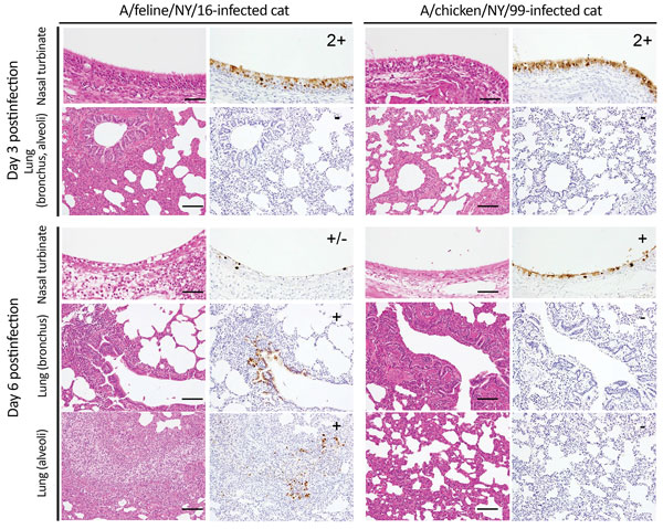
Figure 4. Immunohistochemistry findings in infected cats. Shown are representative sections of nasal turbinates and lungs of cats infected with the indicated viruses on days 3 and 6 postinfection. Three cats per group were infected intranasally with 106 PFU of virus, and tissues were collected on days 3 and 6 postinfection. Type A influenza virus nucleoprotein (NP) was detected by a mouse monoclonal antibody to this protein. For nasal turbinate sections, -: no NP-positive cells, +/−: NP-positive cells detected in 1–2 focal regions, +: NP-positive cells detected in more than two focal regions, 2+: NP-positive cells in large regions. For bronchus and alveolar sections, -: no NP-positive cells, +/−: Up to 5 NP-positive cells, +: Six or more NP-positive cells. NP-positive cells were detected in focal, but not in diffuse bronchial and alveolar sections. For all analyses, the entire sections were evaluated. Scale bars, 50 μm (nasal turbinates), 100 μm (lung).
Main Article
Page created: December 19, 2017
Page updated: December 19, 2017
Page reviewed: December 19, 2017
The conclusions, findings, and opinions expressed by authors contributing to this journal do not necessarily reflect the official position of the U.S. Department of Health and Human Services, the Public Health Service, the Centers for Disease Control and Prevention, or the authors' affiliated institutions. Use of trade names is for identification only and does not imply endorsement by any of the groups named above.