Skip directly to site content Skip directly to page options Skip directly to A-Z link Skip directly to A-Z link Skip directly to A-Z link
Volume 24, Number 1—January 2018
Research

Characterization of a Feline Influenza A(H7N2) Virus

Masato Hatta1, Gongxun Zhong1, Yuwei Gao1, Noriko Nakajima1, Shufang Fan1, Shiho Chiba, Kathleen M. Deering, Mutsumi Ito, Masaki Imai, Maki Kiso, Sumiho Nakatsu, Tiago J. Lopes, Andrew J. Thompson, Ryan McBride, David L. Suarez, Catherine A. Macken, Shigeo Sugita, Gabriele Neumann, Hideki Hasegawa, James C. Paulson, Kathy L. Toohey-Kurth, and Yoshihiro KawaokaComments to Author 
Author affiliations: University of Wisconsin–Madison, Madison, Wisconsin, USA (M. Hatta, G. Zhong, Y. Gao, S. Fan, S. Chiba, K.M. Deering, T.J. Lopes, G. Neumann, K.L. Toohey-Kurth, Y. Kawaoka); National Institute of Infectious Diseases, Tokyo, Japan (N. Nakajima, H. Hasegawa); University of Tokyo, Tokyo (M. Ito, M. Imai, M. Kiso, S. Nakatsu, Y. Kawaoka); The Scripps Research Institute, La Jolla, California, USA (A.J. Thompson, R. McBride, J.C. Paulson); US Department of Agriculture, Athens, Georgia, USA (D.L. Suarez); The University of Auckland, Auckland, New Zealand (C. A. Macken); Japan Racing Association, Tochigi, Japan (S. Sugita)

Main Article

Figure 5

Pathology findings in Feline/NY/16-infected cats on day 6 post-infection. A) In nasal cavities, copious amounts of exudate are present comprising numerous degenerating and necrotic neutrophils, cellular debris, proteinaceous fluid, and strands of mucin. The respiratory epithelium covering the nasal turbinates (T) is extensively eroded. The underlying lamina propria appears diffusely bluish-purple due to infiltration by moderate-to-large numbers of histiocytes, neutrophils, lymphocytes, and plasm

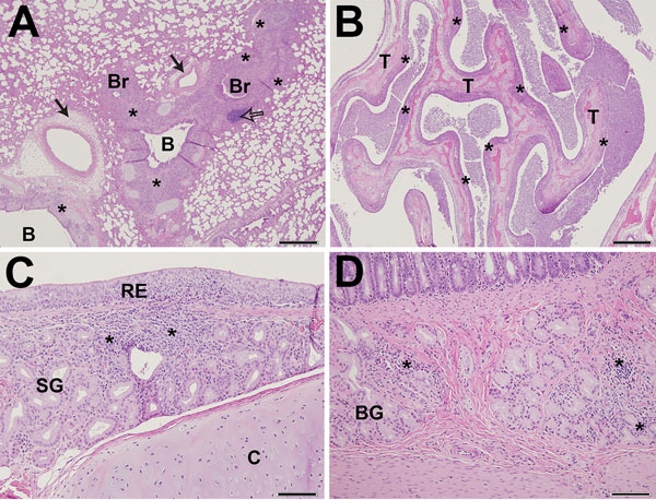
Figure 5. Pathology findings in cats infected with A/feline/NY/16 influenza A(H7N2) virus on day 6 postinfection, New York, NY, USA. A) In lungs, moderately severe histopathologic changes are present in the lower airways. The lamina propria of bronchi (B) and bronchioles (Br) and the surrounding interstitium are infiltrated by numerous histiocytes, lymphocytes, and plasma cells (*), which also extend into and expand neighboring alveolar septa. The infiltrates extend into and expand nearby alveolar septa. The lumina of bronchioles are filled with numerous foamy macrophages, viable and degenerating neutrophils, proteinaceous fluid, and sloughed respiratory epithelial cells. Hyperplasia of bronchiole-associated lymphoid tissue (open arrow) and perivascular edema (solid arrow) are present. Scale bar indicates 500 µm. B) In nasal cavities, copious amounts of exudate are present comprising numerous degenerating and necrotic neutrophils, cellular debris, proteinaceous fluid, and strands of mucin. The respiratory epithelium covering the nasal turbinates (T) is extensively eroded. The underlying lamina propria appears diffusely bluish-purple due to infiltration by moderate-to-large numbers of histiocytes, neutrophils, lymphocytes, and plasma cells (*). Scale bar indicates 500 µm. C) In the trachea, a locally extensive focus of inflammation is present in the tracheal wall. Moderate numbers of histiocytes, lymphocytes, and plasma cells, and a few neutrophils, infiltrate the respiratory epithelium (RE), lamina propria, and submucosa. Submucosal glands (SG) are surrounded by the inflammatory infiltrates and effaced in the areas of heaviest infiltration (*). Tracheal cartilage (C). Scale bar indicates 100 µm. D) In the duodenum, inflammatory cell infiltrates (*) in the submucosa of the duodenum are present between and around Brunner’s glands (BG). Scale bar indicates 100 µm.

Main Article

1These authors contributed equally to this article.

Page created: December 19, 2017
Page updated: December 19, 2017
Page reviewed: December 19, 2017
The conclusions, findings, and opinions expressed by authors contributing to this journal do not necessarily reflect the official position of the U.S. Department of Health and Human Services, the Public Health Service, the Centers for Disease Control and Prevention, or the authors' affiliated institutions. Use of trade names is for identification only and does not imply endorsement by any of the groups named above.
file_external