Volume 24, Number 1—January 2018
Research
Characterization of a Feline Influenza A(H7N2) Virus
Masato Hatta1, Gongxun Zhong1, Yuwei Gao1, Noriko Nakajima1, Shufang Fan1, Shiho Chiba, Kathleen M. Deering, Mutsumi Ito, Masaki Imai, Maki Kiso, Sumiho Nakatsu, Tiago J. Lopes, Andrew J. Thompson, Ryan McBride, David L. Suarez, Catherine A. Macken, Shigeo Sugita, Gabriele Neumann, Hideki Hasegawa, James C. Paulson, Kathy L. Toohey-Kurth, and Yoshihiro Kawaoka
Figure 7
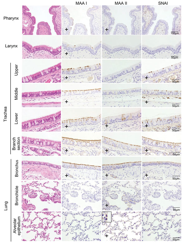
Figure 7. Distribution of α2,3- and α2,6-linked sialosides in the respiratory organs of a cat, New York, NY, USA. The α2,3- and α2,6-linked sialosides in the respiratory organs of a naïve cat were detected with biotinylated Maackia amurensis lectin I or II (MAA I, MAA II) or Sambucus nigra lectin (SNA I), respectively. Inset shows closer view of MAA III binding with alveolar epithelium in the lung. Plus signs (+) indicate that sialosides were detected. Scale bars indicate 50 μm.
1These authors contributed equally to this article.
Page created: December 19, 2017
Page updated: December 19, 2017
Page reviewed: December 19, 2017
The conclusions, findings, and opinions expressed by authors contributing to this journal do not necessarily reflect the official position of the U.S. Department of Health and Human Services, the Public Health Service, the Centers for Disease Control and Prevention, or the authors' affiliated institutions. Use of trade names is for identification only and does not imply endorsement by any of the groups named above.