Volume 29, Number 2—February 2023
Research
Novel Prion Strain as Cause of Chronic Wasting Disease in a Moose, Finland
Figure 2

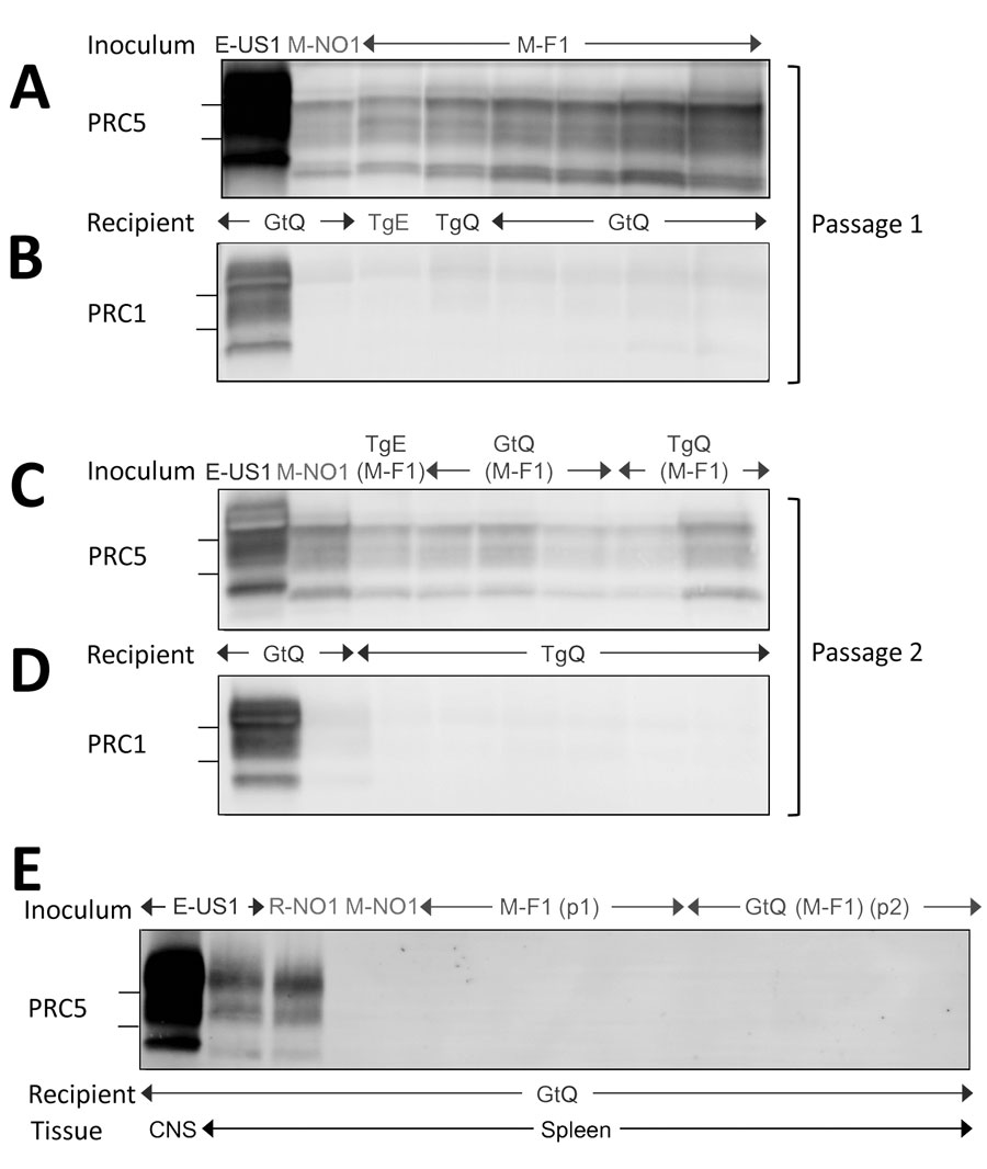
Figure 2. Western blot analyses of cervid prion protein (PrP) scrapie in mice infected with Finland, Norway, and North America chronic wasting disease (CWD) isolates. A, B) Western blots of CNS homogenates from GtQ, TgQ, and TgE mice after primary transmissions E-US1, Norway moose CWD (M-NO1), or Finland moose CWD (M-F1) probed with monoclonal antibody (mAb) PRC5 (A) or PRC1 (B). C–D) Western blots of CNS homogenates of GtQ and TgQ mice infected with E-US1 isolate, Norway isolate M-NO1, and M-F1 passaged through TgE, GtQ, or TgQ, referred to as TgE (M-F1), GtQ (M-F1), and TgQ (M-F1), respectively, probed with mAb PRC5 (C) or PRC1 (D). E) Lane 1, CNS homogenate of GtQ mice infected with E-US1 isolate. Remaining lanes are spleen homogenates from GtQ mice infected with E-US1 isolate, Norway reindeer isolate R-NO1, Norway moose isolate M-NO1, and spleens from infected GtQ mice during primary and secondary transmissions of M-F1. The positions of 25 and 20 kDa molecular weight markers are shown to the left of immunoblots. CNS, central nervous system; E-US1, US elk 1; GtQ, CWD-susceptible gene-targeted mice in which the prion protein coding sequence was replaced with one encoding glutamine at codon 226; M-F1, Finland moose 1; M-NO1, Norway moose 1; PRC1, mAb PCR1; PRC5, mAb PCR5; TgE, transgenic mice expressing cervid PrP with glutamate at residue 226; TgQ, transgenic mice expressing cervid PrP with glutamine at residue 226.