Volume 29, Number 2—February 2023
Research
Novel Prion Strain as Cause of Chronic Wasting Disease in a Moose, Finland
Figure 4
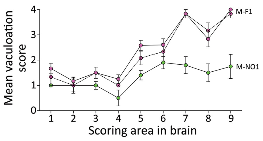
Figure 4. Lesion profiling in GtQ mice infected with Finland and Norway moose chronic wasting disease (CWD) isolates. Lesion profiles in groups of GtQ mice (CWD-susceptible gene-targeted mice in which the prion protein coding sequence was replaced with one encoding glutamine at codon 226) infected with M-F1 (magenta symbols) and Norway moose isolate M-NO1 (green symbols). For M-F1, open circles and solid lines depict primary passage; dotted circles and dashed lines depict second passage. Data points represent the mean +SEM of >5 GtQ mice per group. Brain-scoring areas: medulla (1), cerebellum (2), superior colliculus (3), hypothalamus (4), thalamus (5), hippocampus (6), septum (7), retrosplenial and adjacent motor cortex (8), and cingulate and adjacent motor cortex (9). M-F1, Finland moose 1; M-NO1, Norway moose 1.