Volume 31, Number 12—December 2025
Research
Silent Propagation of Classical Scrapie Prions in Homozygous K222 Transgenic Mice
Natalia Fernández-Borges1, Alba Marín-Moreno1, Juan Carlos Espinosa, Sara Canoyra, Olivier Andréoletti, and Juan María Torres
Figure 3
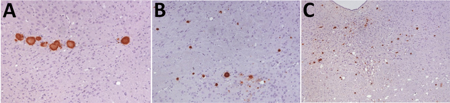
Figure 3. Immunohistochemistry results of brain tissue in study of propagation of classical scrapie prions. Images are of tissue specimens from K222-Tg516 mice inoculated with F10 goat scrapie isolate at second passage. Results are visualized using the Sha31 monoclonal antibody. A) Thalamus specimen. B) Hippocampus specimen. C) Midbrain specimen. Original magnification ×40.
1These first authors contributed equally to this article.
Page created: November 17, 2025
Page updated: January 01, 2026
Page reviewed: January 01, 2026
The conclusions, findings, and opinions expressed by authors contributing to this journal do not necessarily reflect the official position of the U.S. Department of Health and Human Services, the Public Health Service, the Centers for Disease Control and Prevention, or the authors' affiliated institutions. Use of trade names is for identification only and does not imply endorsement by any of the groups named above.