Skip directly to site content Skip directly to page options Skip directly to A-Z link Skip directly to A-Z link Skip directly to A-Z link

Volume 31, Number 12—December 2025

Research

Guinea Pig Model for Lassa Virus Infection of Reproductive Tract and Considerations for Sexual and Vertical Transmission

Josilene N. Seixas, Joy M. Gary1, Stephen R. Welch, Sarah C. Genzer, JoAnn D. Coleman-McCray, Jessica R. Harmon, Christina F. Spiropoulou, Luciana Silva-Flannery, Georgia Ficarra, Elizabeth Lee, Julu Bhatnagar, Jessica R. Spengler, and Jana M. RitterComments to Author 
Author affiliation: Centers for Disease Control and Prevention, Atlanta, Georgia, USA

Main Article

Figure 2

Localization and severity of histopathologic changes and Lassa virus (LASV) detection in reproductive tissues of strain 13/N guinea pigs experimentally infected with LASV strain Josiah or NJ2015 in study of guinea pig model for LASV infection of reproductive tract and considerations for sexual and vertical transmission. Female (A) and male (B) guinea pig reproductive tract anatomy, histopathology, and virus detection (antigen by IHC or viral RNA by ISH) after subcutaneous infection with LASV strain Josiah or NJ-2015 (target dose, 105 focus-forming units). Anatomic localization, severity, and incidence are depicted and delineated by stage of infection: early (<16 dpi), lethal (meeting endpoint criteria at 14–28 dpi), and survivor (41 or 42 dpi). Histopathologic changes (H&E) and viral detection by (IHC or ISH) were scored semiquantitatively for each tissue as absent (0), minimal (1), mild (2), moderate (3), or severe (4). Not all analyses were performed for all animals (Appendix Tables 1–3, https://wwwnc.cdc.gov/EID/article/31/12/25-0396-App1.pdf). H&E, IHC, and ISH scores represent mean severity values for each tissue across all animals tested. An asterisk (*) indicates that only 1 animal was evaluated in the group; gray boxes indicate tissues that were not available or not evaluated. Relative incidence of histopathologic change or virus detection in early, lethal, and survivor cohorts with sufficient group sizes (n >3) is shown in radar plots; only a single male animal in the lethal cohort was excluded, as it was the sole animal evaluated and test results were positive in both mammary gland and skin. H&E, hematoxylin and eosin; IHC, immunohistochemistry; ISH, in situ hybridization.

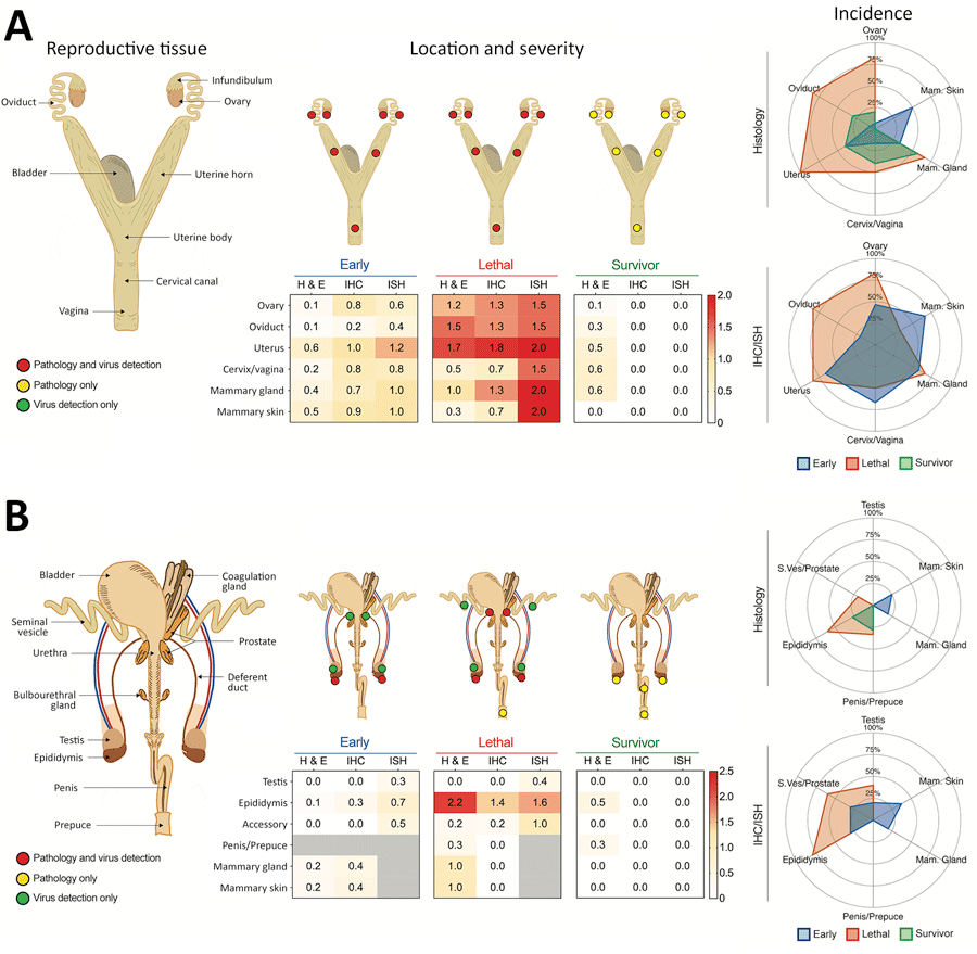
Figure 2. Localization and severity of histopathologic changes and Lassa virus (LASV) detection in reproductive tissues of strain 13/N guinea pigs experimentally infected with LASV strain Josiah or NJ2015 in study of guinea pig model for LASV infection of reproductive tract and considerations for sexual and vertical transmission. Female (A) and male (B) guinea pig reproductive tract anatomy, histopathology, and virus detection (antigen by IHC or viral RNA by ISH) after subcutaneous infection with LASV strain Josiah or NJ-2015 (target dose, 104 focus-forming units). Anatomic localization, severity, and incidence are depicted and delineated by stage of infection: early (<16 dpi), lethal (meeting endpoint criteria at 14–28 dpi), and survivor (41 or 42 dpi). Histopathologic changes (H&E) and viral detection by (IHC or ISH) were scored semiquantitatively for each tissue as absent (0), minimal (1), mild (2), moderate (3), or severe (4). Not all analyses were performed for all animals (Appendix Tables 1–3, https://wwwnc.cdc.gov/EID/article/31/12/25-0396-App1.pdf). H&E, IHC, and ISH scores represent mean severity values for each tissue across all animals tested. An asterisk (*) indicates that only 1 animal was evaluated in the group; gray boxes indicate tissues that were not available or not evaluated. Relative incidence of histopathologic change or virus detection in early, lethal, and survivor cohorts with sufficient group sizes (n >3) is shown in radar plots; only a single male animal in the lethal cohort was excluded, as it was the sole animal evaluated and test results were positive in both mammary gland and skin. H&E, hematoxylin and eosin; IHC, immunohistochemistry; ISH, in situ hybridization.

Main Article

1Current affiliation: StageBio, Frederick, Maryland, USA.

Page created: November 25, 2025
Page updated: January 01, 2026
Page reviewed: January 01, 2026
The conclusions, findings, and opinions expressed by authors contributing to this journal do not necessarily reflect the official position of the U.S. Department of Health and Human Services, the Public Health Service, the Centers for Disease Control and Prevention, or the authors' affiliated institutions. Use of trade names is for identification only and does not imply endorsement by any of the groups named above.
file_external