Skip directly to site content Skip directly to page options Skip directly to A-Z link Skip directly to A-Z link Skip directly to A-Z link

Volume 31, Number 12—December 2025

Research

Guinea Pig Model for Lassa Virus Infection of Reproductive Tract and Considerations for Sexual and Vertical Transmission

Josilene N. Seixas, Joy M. Gary1, Stephen R. Welch, Sarah C. Genzer, JoAnn D. Coleman-McCray, Jessica R. Harmon, Christina F. Spiropoulou, Luciana Silva-Flannery, Georgia Ficarra, Elizabeth Lee, Julu Bhatnagar, Jessica R. Spengler, and Jana M. RitterComments to Author 
Author affiliation: Centers for Disease Control and Prevention, Atlanta, Georgia, USA

Main Article

Figure 7

Histopathologic findings in reproductive tissues from strain 13/N guinea pigs serially euthanized 4–16 days postinfection (dpi) after Lassa virus strain Josiah infection in study of guinea pig model for infection of reproductive tract and considerations for sexual and vertical transmission. A) Perivascular mononuclear infiltrates (arrow) in oviduct (12 dpi). B) Perivascular (arrow) and interstitial (asterisk) lymphoplasmacytic infiltrates within the myometrium in uterus (16 dpi). C) Interstitial and perivascular lymphoplasmacytic infiltrates (arrows), with rare single cell necrosis of the tubular epithelial cells (asterisk) in epididymis (16 dpi). D) Mild perivascular and vascular inflammation (arrow) and interstitial edema (asterisk) in epididymis (16 dpi). Hematoxylin and eosin stain. Original magnifications ×40 (panels A, B, D), ×20 (panel C).

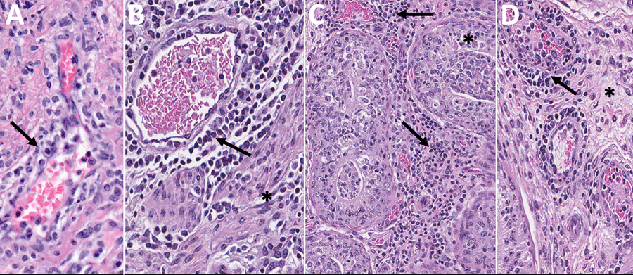
Figure 7. Histopathologic findings in reproductive tissues from strain 13/N guinea pigs serially euthanized 4–16 days postinfection (dpi) after Lassa virus strain Josiah infection in study of guinea pig model for infection of reproductive tract and considerations for sexual and vertical transmission. A) Perivascular mononuclear infiltrates (arrow) in oviduct (12 dpi). B) Perivascular (arrow) and interstitial (asterisk) lymphoplasmacytic infiltrates within the myometrium in uterus (16 dpi). C) Interstitial and perivascular lymphoplasmacytic infiltrates (arrows), with rare single cell necrosis of the tubular epithelial cells (asterisk) in epididymis (16 dpi). D) Mild perivascular and vascular inflammation (arrow) and interstitial edema (asterisk) in epididymis (16 dpi). Hematoxylin and eosin stain. Original magnifications ×40 (panels A, B, D), ×20 (panel C).

Main Article

1Current affiliation: StageBio, Frederick, Maryland, USA.

Page created: November 25, 2025
Page updated: January 01, 2026
Page reviewed: January 01, 2026
The conclusions, findings, and opinions expressed by authors contributing to this journal do not necessarily reflect the official position of the U.S. Department of Health and Human Services, the Public Health Service, the Centers for Disease Control and Prevention, or the authors' affiliated institutions. Use of trade names is for identification only and does not imply endorsement by any of the groups named above.
file_external