Skip directly to site content Skip directly to page options Skip directly to A-Z link Skip directly to A-Z link Skip directly to A-Z link

Volume 31, Number 12—December 2025

Research

Guinea Pig Model for Lassa Virus Infection of Reproductive Tract and Considerations for Sexual and Vertical Transmission

Josilene N. Seixas, Joy M. Gary1, Stephen R. Welch, Sarah C. Genzer, JoAnn D. Coleman-McCray, Jessica R. Harmon, Christina F. Spiropoulou, Luciana Silva-Flannery, Georgia Ficarra, Elizabeth Lee, Julu Bhatnagar, Jessica R. Spengler, and Jana M. RitterComments to Author 
Author affiliation: Centers for Disease Control and Prevention, Atlanta, Georgia, USA

Main Article

Figure 8

Detection of viral antigens and RNA by immunohistochemistry (IHC) and in situ hybridization (ISH) in reproductive tissues from strain 13/N guinea pigs serially euthanized 4–16 dpi after LASV strain Josiah infection in study of guinea pig model for infection of reproductive tract and considerations for sexual and vertical transmission. IHC and ISH chromogens are red. Panels A–G depict samples from female guinea pigs; panels H–L depict samples from male guinea pigs. A) Uterus (4 dpi), ISH. Rare staining in the endometrial stroma (arrow), without inflammation. B) Endocervix (4 dpi), ISH. Staining of the apical surface of an endocervical epithelial cell (asterisk) and beneath the basilar epithelium (arrow), without inflammation. C) Ovary (8 dpi), IHC. Staining in granulosa lutein cells (arrow) of a corpus luteum without inflammation. D) Uterus (8dpi), IHC. Staining in endometrial stromal cells (arrow) without inflammation. E) Ovary (12 dpi), ISH. Staining in theca interstitial cells (asterisk) and endothelium (arrow). F) Uterus (12 dpi), IHC. Endometrial stromal lymphoplasmacytic infiltrates and staining in endometrial stromal (asterisk) and epithelial (arrows) cells. G) Ovary (16 dpi), IHC. Staining in granulosa cells (asterisk) and endothelium (arrow). H) Epididymis (8 dpi), IHC. Staining in peritubular interstitial mesenchymal cells (arrow). I) Seminal vesicle (12 dpi), ISH. Staining in the wall of a small interstitial vessel (arrow). J) Testis (12 dpi), ISH. Rare granular staining of viral RNA in the interstitium (white arrow) and in a vascular lumen (arrow). K, L) Epididymis (16 dpi), ISH. Extensive staining in tubular epithelium (arrows) and within tubular lumens, including spermatozoa (asterisk). Original magnifications ×40 (panels A, C, D, F, G, H, J, L), ×63 (panels B, I), ×10 (panel E), ×4 (panel K).

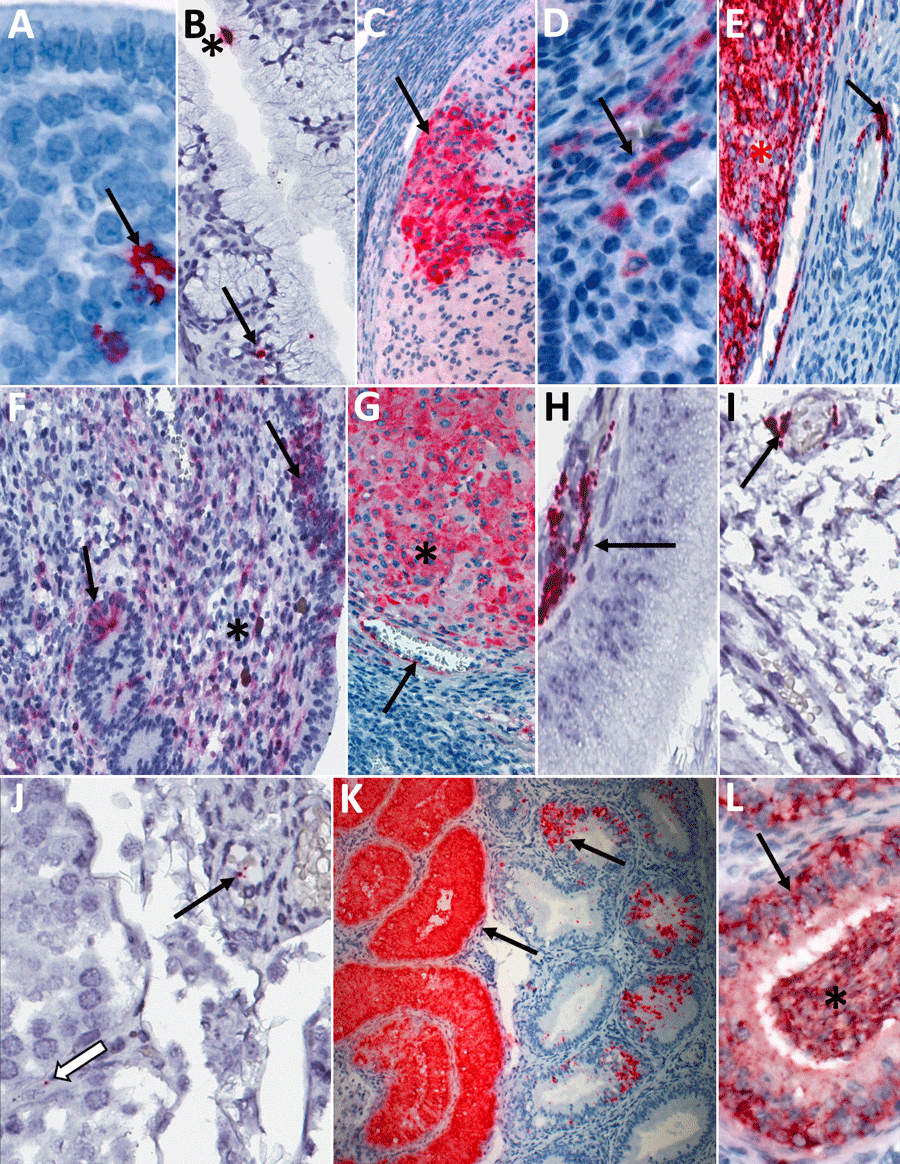
Figure 8. Detection of viral antigens and RNA by immunohistochemistry (IHC) and in situ hybridization (ISH) in reproductive tissues from strain 13/N guinea pigs serially euthanized 4–16 dpi after LASV strain Josiah infection in study of guinea pig model for infection of reproductive tract and considerations for sexual and vertical transmission. IHC and ISH chromogens are red. Panels A–G depict samples from female guinea pigs; panels H–L depict samples from male guinea pigs. A) Uterus (4 dpi), ISH. Rare staining in the endometrial stroma (arrow), without inflammation. B) Endocervix (4 dpi), ISH. Staining of the apical surface of an endocervical epithelial cell (asterisk) and beneath the basilar epithelium (arrow), without inflammation. C) Ovary (8 dpi), IHC. Staining in granulosa lutein cells (arrow) of a corpus luteum without inflammation. D) Uterus (8dpi), IHC. Staining in endometrial stromal cells (arrow) without inflammation. E) Ovary (12 dpi), ISH. Staining in theca interstitial cells (asterisk) and endothelium (arrow). F) Uterus (12 dpi), IHC. Endometrial stromal lymphoplasmacytic infiltrates and staining in endometrial stromal (asterisk) and epithelial (arrows) cells. G) Ovary (16 dpi), IHC. Staining in granulosa cells (asterisk) and endothelium (arrow). H) Epididymis (8 dpi), IHC. Staining in peritubular interstitial mesenchymal cells (arrow). I) Seminal vesicle (12 dpi), ISH. Staining in the wall of a small interstitial vessel (arrow). J) Testis (12 dpi), ISH. Rare granular staining of viral RNA in the interstitium (white arrow) and in a vascular lumen (arrow). K, L) Epididymis (16 dpi), ISH. Extensive staining in tubular epithelium (arrows) and within tubular lumens, including spermatozoa (asterisk). Original magnifications ×40 (panels A, C, D, F, G, H, J, L), ×63 (panels B, I), ×10 (panel E), ×4 (panel K).

Main Article

1Current affiliation: StageBio, Frederick, Maryland, USA.

Page created: November 25, 2025
Page updated: January 01, 2026
Page reviewed: January 01, 2026
The conclusions, findings, and opinions expressed by authors contributing to this journal do not necessarily reflect the official position of the U.S. Department of Health and Human Services, the Public Health Service, the Centers for Disease Control and Prevention, or the authors' affiliated institutions. Use of trade names is for identification only and does not imply endorsement by any of the groups named above.
file_external