Skip directly to site content Skip directly to page options Skip directly to A-Z link Skip directly to A-Z link Skip directly to A-Z link

Volume 31, Number 12—December 2025

Research

Guinea Pig Model for Lassa Virus Infection of Reproductive Tract and Considerations for Sexual and Vertical Transmission

Josilene N. Seixas, Joy M. Gary1, Stephen R. Welch, Sarah C. Genzer, JoAnn D. Coleman-McCray, Jessica R. Harmon, Christina F. Spiropoulou, Luciana Silva-Flannery, Georgia Ficarra, Elizabeth Lee, Julu Bhatnagar, Jessica R. Spengler, and Jana M. RitterComments to Author 
Author affiliation: Centers for Disease Control and Prevention, Atlanta, Georgia, USA

Main Article

Figure 9

Histopathologic findings in reproductive tissues from strain 13/N guinea pig survivors of Lassa virus strain Josiah infection at 42 days postinfection (dpi) in study of guinea pig model for Lassa virus infection of reproductive tract and considerations for sexual and vertical transmission. Panels A–D depict samples from female guinea pigs; panels E–F depict male samples from male guinea pigs. A) Ovary (42 dpi) with lymphoplasmacytic inflammation in the stroma (asterisk). B) Oviduct (42 dpi) with focal perivascular and interstitial inflammation (arrow). C) Uterus (42 dpi) with endometrial stromal lymphoplasmacytic inflammation (asterisk) around glands. D) Vagina (42 dpi) with lymphocytic inflammation (arrows) around vessels in the vaginal wall. E) Epididymis (42 dpi) with dense perivascular lymphoplasmacytic inflammation around a vessel (white arrow) and heterophils within tubular lumens (black arrows). F) Penile connective tissue (42 dpi) with perivascular lymphocytic inflammation (arrow). Hematoxylin and eosin stain. Original magnification ×40 (panels A–C, E, F), ×10 (panel D).

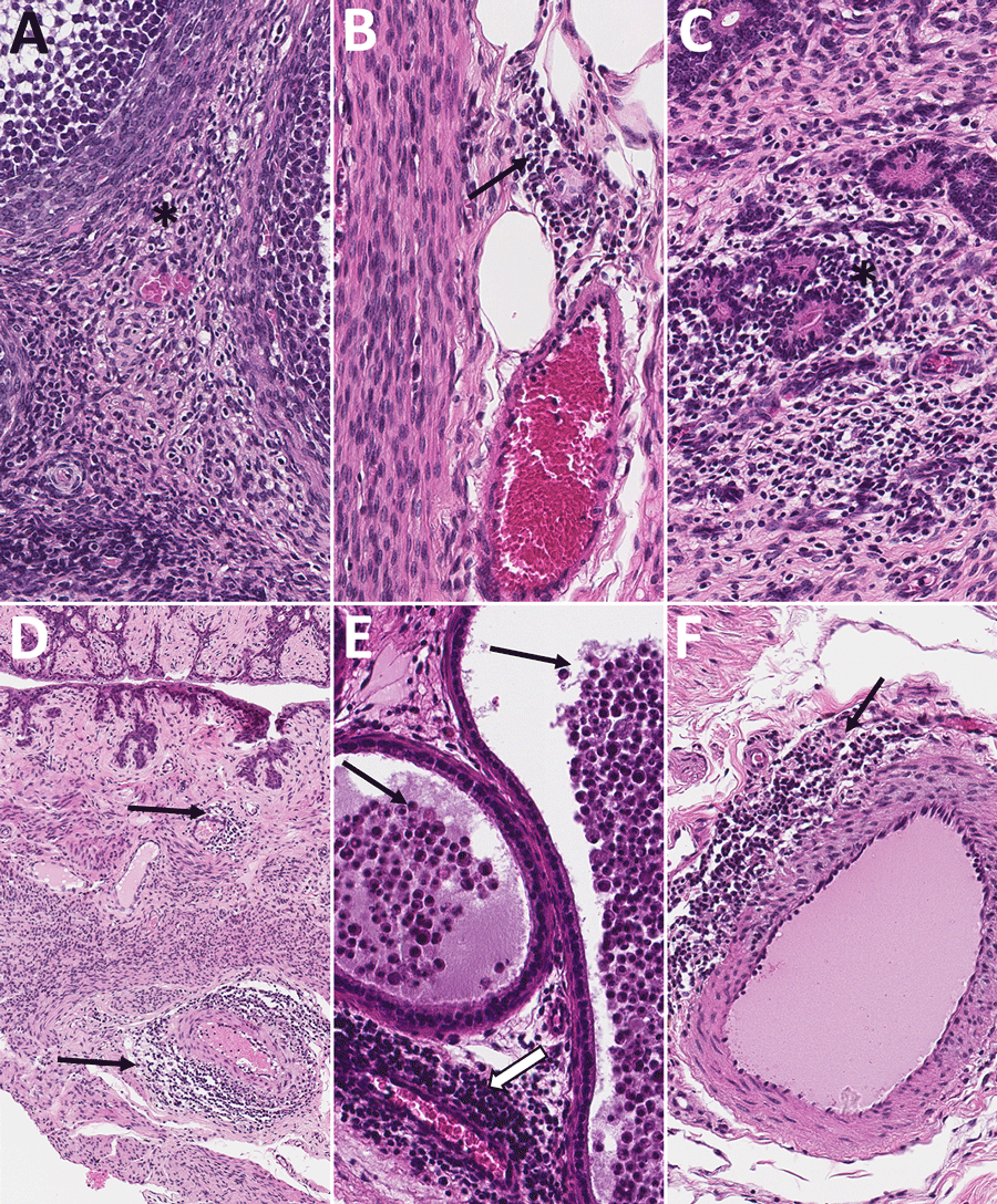
Figure 9. Histopathologic findings in reproductive tissues from strain 13/N guinea pig survivors of Lassa virus strain Josiah infection at 42 days postinfection (dpi) in study of guinea pig model for Lassa virus infection of reproductive tract and considerations for sexual and vertical transmission. Panels A–D depict samples from female guinea pigs; panels E–F depict male samples from male guinea pigs. A) Ovary (42 dpi) with lymphoplasmacytic inflammation in the stroma (asterisk). B) Oviduct (42 dpi) with focal perivascular and interstitial inflammation (arrow). C) Uterus (42 dpi) with endometrial stromal lymphoplasmacytic inflammation (asterisk) around glands. D) Vagina (42 dpi) with lymphocytic inflammation (arrows) around vessels in the vaginal wall. E) Epididymis (42 dpi) with dense perivascular lymphoplasmacytic inflammation around a vessel (white arrow) and heterophils within tubular lumens (black arrows). F) Penile connective tissue (42 dpi) with perivascular lymphocytic inflammation (arrow). Hematoxylin and eosin stain. Original magnification ×40 (panels A–C, E, F), ×10 (panel D).

Main Article

1Current affiliation: StageBio, Frederick, Maryland, USA.

Page created: November 25, 2025
Page updated: January 01, 2026
Page reviewed: January 01, 2026
The conclusions, findings, and opinions expressed by authors contributing to this journal do not necessarily reflect the official position of the U.S. Department of Health and Human Services, the Public Health Service, the Centers for Disease Control and Prevention, or the authors' affiliated institutions. Use of trade names is for identification only and does not imply endorsement by any of the groups named above.
file_external