Volume 31, Number 12—December 2025
Research
Guinea Pig Model for Lassa Virus Infection of Reproductive Tract and Considerations for Sexual and Vertical Transmission
Figure 5
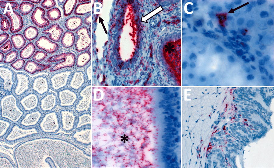
Figure 5. Detection of Lassa virus (LASV) antigens and RNA in reproductive tissues by immunohistochemistry (IHC) and in situ hybridization (ISH) in strain 13/N male guinea pigs lethally infected with LASV strain Josiah in study of guinea pig model for LASV infection of reproductive tract and considerations for sexual and vertical transmission. A) Epididymis (18 dpi), ISH. Regionally extensive staining in epididymal epithelial cells. B) Epididymis (26 dpi), ISH. Staining in interstitial mesenchymal cells, endothelium (arrow), tubular epithelium (white arrow), and intratubular inflammatory cells (asterisk). C) Testis (18 dpi), ISH. Focal intracellular staining in the interstitium (arrow). D) Epididymis (23 dpi), ISH. Intraluminal staining including spermatozoa (asterisk). E) Seminal vesicle (17 dpi), ISH. Focal staining in subepithelial stromal cells. Original magnifications ×5 (panel A), ×40 (panels B, D), ×63 (panel C), ×20 (panel E).
1Current affiliation: StageBio, Frederick, Maryland, USA.