Influenza A(H5N1) Immune Response among Ferrets with Influenza A(H1N1)pdm09 Immunity
Valerie Le Sage

, Bailee D. Werner, Grace A. Merrbach, Sarah E. Petnuch, Aoife K. O’Connell, Holly C. Simmons, Kevin R. McCarthy, Douglas S. Reed, Louise H. Moncla, Disha Bhavsar, Florian Krammer, Nicholas A. Crossland, Anita K. McElroy, W. Paul Duprex, and Seema S. Lakdawala
Author affiliation: Author affiliations: University of Pittsburgh, Pittsburgh, Pennsylvania, USA (V. Le Sage, B.D. Werner, G.A. Merrbach, S.E. Petnuch, H.C. Simmons, K.R. McCarthy, D.S. Reed, L.H. Moncla, A.K. McElroy, W.P. Duprex); Boston University, Boston, Massachusetts, USA (A.K. O’Connell, N.A. Crossland); Icahn School of Medicine at Mount Sinai, New York, New York, USA (D. Bhavsar, F. Krammer); Medical University of Vienna, Vienna, Austria (F. Krammer); Boston University Chobanian & Avedisian School of Medicine, Boston, Massachusetts, USA (N.A. Crossland); Emory University, Atlanta, Georgia, USA (S.S. Lakdawala)
Main Article
Figure 4
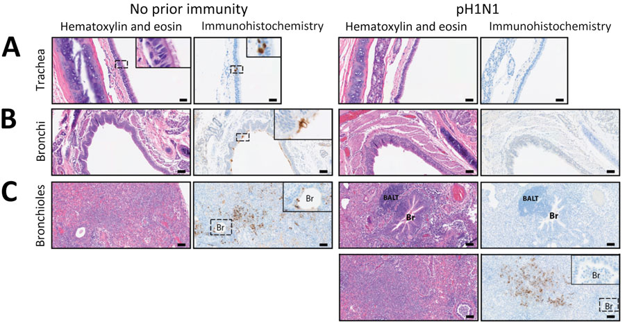
Figure 4. Hematoxylin and eosin–stained and immunohistochemistry tissue samples from a study of influenza A(H5N1) immune response among ferrets with pH1N1 immunity. A) Tracheal tissue. Scale bars indicate 20 mm; inset shows magnification ×400. B) Bronchial tissue. Scale bars indicate 50 mm; inset shows magnification ×200. C) Bronchiole tissue. Scale bars indicate 50 mm; inset shows magnification ×200. Ferrets with no prior immunity (left panels) or existing influenza A(H1N1)pdm09 immunity (right panels) were infected with 104 50% tissue culture infectious dose of H5N1 strain A/dairy cattle/Texas/24-008749-001/2024(H5N1) and humanely euthanized 3 days postinfection. Images show hematoxylin and eosin stained (purple) tissues and immunohistochemistry of influenza A nucleoprotein (blue). Dotted squares indicate areas that are magnified within the inset panel in tissues from ferrets with no prior immunity versus pH1N1-immune ferrets. BALT, bronchus-associated lymphoid tissue; Br, bronchiole; pH1N1, influenza A(H1N1)pdm09.
Main Article
Page created: January 21, 2025
Page updated: February 28, 2025
Page reviewed: February 28, 2025
The conclusions, findings, and opinions expressed by authors contributing to this journal do not necessarily reflect the official position of the U.S. Department of Health and Human Services, the Public Health Service, the Centers for Disease Control and Prevention, or the authors' affiliated institutions. Use of trade names is for identification only and does not imply endorsement by any of the groups named above.