Peromyscus spp. Deer Mice as Rodent Model of Acute Leptospirosis
Ellie J. Putz

, Claire B. Andreasen, Paola Boggiatto, Mitchell V. Palmer, Luis G.V. Fernandes, Bienvenido W. Tibbs-Cortes, Judith A. Stasko, Camila Hamond, Steven C. Olsen, and Jarlath E. Nally
Author affiliation: National Animal Disease Center, US Department of Agriculture Agricultural Research Service, Ames, Iowa, USA (E.J. Putz, P. Boggiatto, M.V. Palmer, L.G.V. Fernandes, B.W. Tibbs-Cortes, J.A. Stasko, S.C. Olsen, J.E. Nally); Iowa State University College of Veterinary Medicine, Ames (C.B. Andreasen); National Veterinary Services Laboratories, US Department of Agriculture Animal and Plant Health Inspection Service, Ames (C. Hamond)
Main Article
Figure 6
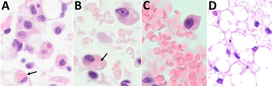
Figure 6. Histological examination of omental tissue from Peromyscus deer mice in study of the species as rodent model of acute leptospirosis. A) Leptospira borgpetersenii strain LR131–challenged deer mouse (original magnification ×40); B) foamy macrophages with intracytoplasmic erythrocytes and clear lipid vacuoles in sample taken from LR131-challenged deer mouse (original magnification ×100); C) foamy macrophage within vessel lumen and foamy macrophage, also present in peripheral blood L. interrogans strain LAD1–challenged deer mouse (original magnification ×100); D) omentum sample from noninfected deer mouse (original magnification ×40). Black arrows indicate possible foamy macrophages/macrophages with intracytoplasmic erythrocytes (erythrophagocytosis).
Main Article
Page created: May 22, 2025
Page updated: June 25, 2025
Page reviewed: June 25, 2025
The conclusions, findings, and opinions expressed by authors contributing to this journal do not necessarily reflect the official position of the U.S. Department of Health and Human Services, the Public Health Service, the Centers for Disease Control and Prevention, or the authors' affiliated institutions. Use of trade names is for identification only and does not imply endorsement by any of the groups named above.