Skip directly to site content Skip directly to page options Skip directly to A-Z link Skip directly to A-Z link Skip directly to A-Z link

Disclaimer: Early release articles are not considered as final versions. Any changes will be reflected in the online version in the month the article is officially released.

Volume 31, Number 7—July 2025

Research

Peromyscus spp. Deer Mice as Rodent Model of Acute Leptospirosis

Ellie J. PutzComments to Author , Claire B. Andreasen, Paola Boggiatto, Mitchell V. Palmer, Luis G.V. Fernandes, Bienvenido W. Tibbs-Cortes, Judith A. Stasko, Camila Hamond, Steven C. Olsen, and Jarlath E. Nally
Author affiliation: National Animal Disease Center, US Department of Agriculture Agricultural Research Service, Ames, Iowa, USA (E.J. Putz, P. Boggiatto, M.V. Palmer, L.G.V. Fernandes, B.W. Tibbs-Cortes, J.A. Stasko, S.C. Olsen, J.E. Nally); Iowa State University College of Veterinary Medicine, Ames (C.B. Andreasen); National Veterinary Services Laboratories, US Department of Agriculture Animal and Plant Health Inspection Service, Ames (C. Hamond)

Main Article

Figure 8

Microscopic and immunohistochemical examination of liver samples from challenged and control deer mice in study of Peromyscus species deer mice as rodent model of acute leptospirosis. A–C) Samples from LR131-challenged deer mice showing minimal change and intravascular neutrophils. The liver had intact architecture, minimal hepatocyte change, and some areas of vascular intrasinusoidal neutrophils. The presence of neutrophils may represent peripheral blood versus recruitment to the liver. A) Hematoxylin and eosin–stained tissue (A, original magnification ×20; B, original magnification ×40); (C) LipL32 immunohistochemistry-labeled liver (original magnification ×20). D–F) Samples from LAD1-challenged deer mice showing acute diffuse purulent hepatitis and necrosis. There was marked disruption of hepatic architecture with hepatocyte dissociation, diffuse neutrophilic (purulent) inflammation, and hepatocyte necrosis. Necrosis of hepatocytes resulted in confluent midzonal and centrilobular necrosis, sometimes bridging, with hemorrhage. The necrosis resulted in tissue fragility. D) Hematoxylin and eosin–stained tissue (A, original magnification ×20; B, original magnification ×40); F) LipL32 immunohistochemistry-labeled liver (original magnification ×20). G–I) Sample of noninfected control animal liver. G) Hematoxylin and eosin–stained tissue (A, original magnification ×20; B, original magnification ×40); I) LipL32 immunohistochemistry negative (original magnification ×20).

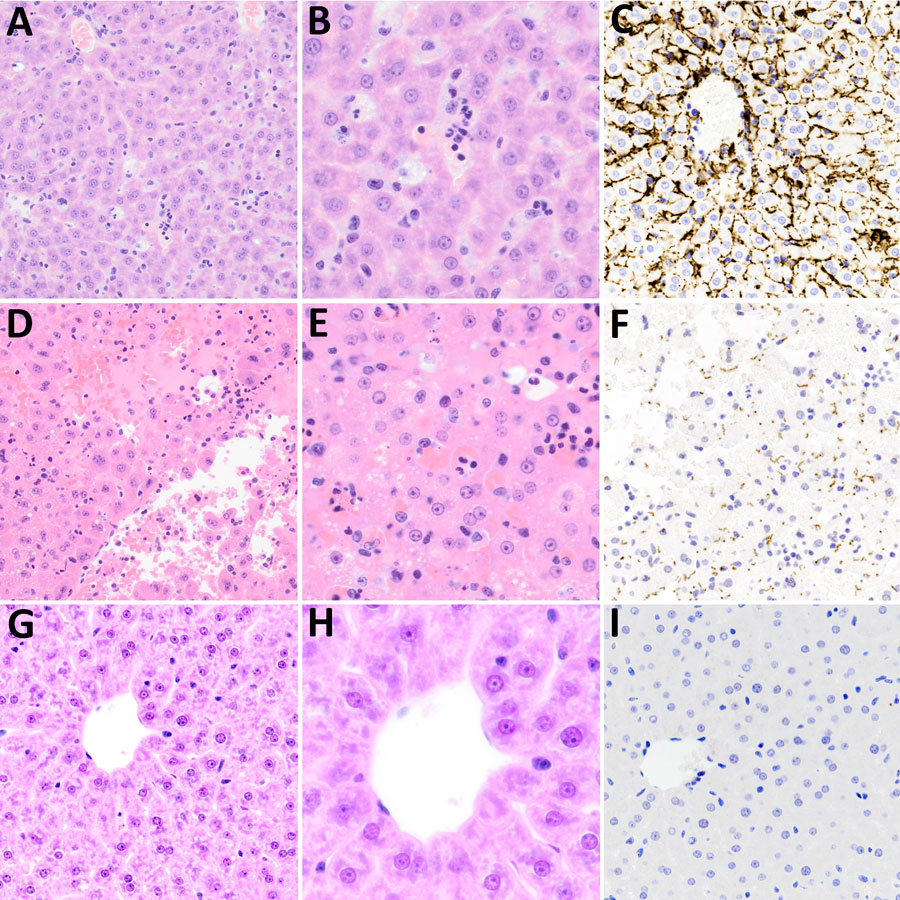
Figure 8. Microscopic and immunohistochemical examination of liver samples from challenged and control deer mice in study of Peromyscus species deer mice as rodent model of acute leptospirosis. A–C) Samples from LR131-challenged deer mice showing minimal change and intravascular neutrophils. The liver had intact architecture, minimal hepatocyte change, and some areas of vascular intrasinusoidal neutrophils. The presence of neutrophils may represent peripheral blood versus recruitment to the liver. A) Hematoxylin and eosin–stained tissue (A, original magnification ×20; B, original magnification ×40); (C) LipL32 immunohistochemistry-labeled liver (original magnification ×20). D–F) Samples from LAD1-challenged deer mice showing acute diffuse purulent hepatitis and necrosis. There was marked disruption of hepatic architecture with hepatocyte dissociation, diffuse neutrophilic (purulent) inflammation, and hepatocyte necrosis. Necrosis of hepatocytes resulted in confluent midzonal and centrilobular necrosis, sometimes bridging, with hemorrhage. The necrosis resulted in tissue fragility. D) Hematoxylin and eosin–stained tissue (A, original magnification ×20; B, original magnification ×40); F) LipL32 immunohistochemistry-labeled liver (original magnification ×20). G–I) Sample of noninfected control animal liver. G) Hematoxylin and eosin–stained tissue (A, original magnification ×20; B, original magnification ×40); I) LipL32 immunohistochemistry negative (original magnification ×20).

Main Article

Page created: May 22, 2025
Page updated: June 12, 2025
Page reviewed: June 12, 2025
The conclusions, findings, and opinions expressed by authors contributing to this journal do not necessarily reflect the official position of the U.S. Department of Health and Human Services, the Public Health Service, the Centers for Disease Control and Prevention, or the authors' affiliated institutions. Use of trade names is for identification only and does not imply endorsement by any of the groups named above.
file_external