Volume 31, Number 7—July 2025
Research
Peromyscus spp. Deer Mice as Rodent Model of Acute Leptospirosis
Ellie J. Putz , Claire B. Andreasen, Paola Boggiatto, Mitchell V. Palmer, Luis G.V. Fernandes, Bienvenido W. Tibbs-Cortes, Judith A. Stasko, Camila Hamond, Steven C. Olsen, and Jarlath E. Nally
Figure 4
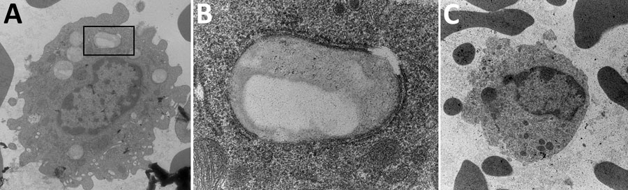
Figure 4. Transmission electron microscopy presentation of whole blood indicating foamy macrophages from infected mice in study of Peromyscus species deer mice as rodent model of acute leptospirosis. A) Image of foamy macrophages with membrane-bound lipid droplet in black box; original magnification ×6,800. B) Close-up of membrane-bound lipid droplet from box in panel A (original magnification ×49,000). C) Noninfected control monocyte (original magnification ×6,800).
Page created: May 22, 2025
Page updated: June 25, 2025
Page reviewed: June 25, 2025
The conclusions, findings, and opinions expressed by authors contributing to this journal do not necessarily reflect the official position of the U.S. Department of Health and Human Services, the Public Health Service, the Centers for Disease Control and Prevention, or the authors' affiliated institutions. Use of trade names is for identification only and does not imply endorsement by any of the groups named above.