Volume 32, Number 9—September 2026
Research
Clustering and Source Association of Clinical and Nonclinical Listeria monocytogenes Isolates, New York, USA, 2000–20211


Cite This Article
Citation for Media
Abstract
We analyzed whole-genome sequencing data for 1,046 human clinical and 1,332 nonclinical Listeria monocytogenes isolates collected across New York, USA, during 2000–2021. Several hypervirulent clonal complexes (CCs) were significantly associated with clinical isolates, and several hypovirulent CCs were associated with nonclinical isolates. Specific CCs also showed association with specific food categories (e.g., processed meat); specific genetic markers (e.g., inlA premature stop codons) were also significantly associated with processed meat isolates. Analysis of clusters that contained food isolates, as well as subsequently identified clinical isolates, showed that time of isolation between food isolates and clinical isolates was significantly shorter for produce isolates than for isolates from meat, dairy, or fish. This finding suggests unique transmission pathways for produce, which might reflect short shelf life or limited L. monocytogenes persistence (e.g., in agricultural environments). This study highlights new opportunities for use of whole-genome sequencing to improve outbreak investigations and source attribution.
Listeria monocytogenes is a leading cause of foodborne illness–related deaths in the United States; an estimated 1,250 human illnesses and 172 deaths occurred annually during 2016–2019 (1). L. monocytogenes is frequently isolated from dairy, ready-to-eat meats, seafood, fruits, and vegetables, and it can persist in food processing environments (2), leading to recurrent contamination of finished products. Hence, robust surveillance is critical for protecting vulnerable populations and rapid detection of contamination sources.
Multilocus sequence typing (MLST) is widely used to classify L. monocytogenes into clonal complexes (CCs), and certain CCs are suggested as presenting higher and lower virulence (3). CC1, CC4, and CC6 are considered hypervirulent and are frequently associated with human listeriosis cases (4). Conversely, CC9 and CC121 are frequently isolated from food or food-processing environments and are considered hypovirulent CCs (4,5). Hypovirulent CCs often carry adaptations that promote environmental persistence (e.g., stress tolerance genes) but might have reduced ability to cause invasive disease (6). Characterizing the distribution of CCs across clinical and nonclinical isolates is key for identifying contamination reservoirs and understanding the public health relevance of isolates found in foods. Distinguishing hypervirulent strains from those mainly associated with the environment can help prioritize risk management and improve listeriosis prevention strategies (7). In this study, we analyzed 2,371 clinical and nonclinical L. monocytogenes isolates collected across the state of New York (NY), USA, during 2000–2021 to compare the CC distributions among clinical and nonclinical isolates, examine associations between CCs and food categories, and evaluate the time between food isolate collection and subsequent detection of a genetically-related clinical isolate using single-nucleotide polymorphism (SNP)–based thresholds. The Wadsworth Center and Cornell University Institutional Review Boards reviewed this study and determined it to be exempt from human research (submission no. 03-037 and IRB0150826).
Detailed materials and methods are provided in the Appendix. In brief, we performed whole-genome sequencing (WGS) on clinical (n = 1,046) and nonclinical (n = 1,332) L. monocytogenes isolates collected across NY, as previously described (8). We deduplicated clinical isolates from the same patient or mother–infant pairs, resulting in 1,006 clinical isolates. Likewise, we deduplicated multiple nonclinical isolates from the same sampling event, resulting in 1,089 nonclinical isolates.
We used whole-genome SNP distances to assess the genetic relatedness among isolates, as previously described (8). We used genetic relatedness of clinical and nonclinical isolates to define 3 types of isolate grouping. When only clinical isolates were involved, we used the term clustered, and when clinical and nonclinical isolates were involved, we used the term linked. Hence, the term linked was used to describe genomic similarity between clinical and nonclinical isolates on the basis of SNP thresholds, rather than confirmed epidemiologic associations. Therefore, NY clinical isolates were considered clustered if clustered with >1 other NY clinical isolate by <20 SNPs (9–11), NY nonclinical isolates were considered cluster-linked if linked by <50 SNPs with >1 clustered NY clinical isolate (12), and NY clinical and nonclinical isolates were considered sporadic-linked if a NY clinical isolate was not clustered with any other NY clinical isolate by <20 SNPs but was linked with >1 NY nonclinical isolate by <50 SNPs. The terms clustered, cluster-linked, and sporadic-linked refer only to genetic relatedness on the basis of SNP thresholds, without interpretation of epidemiologic data. Isolates not meeting those criteria were considered unclustered NY clinical isolates and unlinked NY nonclinical isolates.
CC Distributions among Clinical and Nonclinical Isolates
Figure 1
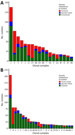
Figure 1. Distribution of Listeria monocytogenes CCs by grouping status in terms of genetical relatedness by single-nucleotide polymorphism (SNP) distances in study of clustering and source association of clinical and nonclinical ...
Figure 2
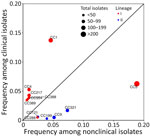
Figure 2. Comparison of the relative frequency of Listeria monocytogenes CCs among clinical and nonclinical isolates in study of clustering and source association of clinical and nonclinical L. monocytogenes...
Among clinical isolates, CC1 (n = 138), CC6 (n = 77), CC5 (n = 63), and CC4 (n = 53) were the most prevalent CCs (Figure 1, panel A). Among nonclinical isolates, CC5 (n = 205), CC321 (n = 80), CC6 (n = 80), and CC2 (n = 69) were the most prevalent CCs (Figure 1, panel B). CC1, CC4, CC217, CC388, CC389, and CC554 were significantly overrepresented among clinical isolates, whereas CC5, CC9, CC121, CC199, CC288, and CC321 were significantly overrepresented among nonclinical isolates (Table 1; Figure 2).
On the basis of their grouping patterns within NY isolates, 318 (31.6%) clinical isolates were clustered, 79 (7.9%) were sporadic-linked, and 609 (60.5%) were unclustered (Figure 1, panel A). Among nonclinical isolates, 236 (21.7%) were classified as cluster-linked, 107 (9.8%) were sporadic-linked, and 746 (68.5%) were unlinked (Figure 1, panel B). Most CC321 nonclinical isolates were cluster-linked, whereas CC6, CC2, CC9, and CC121 nonclinical isolates were predominantly unlinked (e.g., 59 out of 61 CC9 nonclinical isolates were unlinked).
Association of CCs with Food Categories in NY
Figure 3
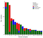
Figure 3. Distribution of Listeria monocytogenesclonal complexe (CCs) by grouping status in terms of genetical relatedness by single-nucleotide polymorphism (SNP) distances among food isolates in study of clustering and source...
The 313 food isolates (28.7% of nonclinical isolates) came from processed meat (n = 93), processed fish (n = 72), raw dairy (n = 47), raw fish (n = 22), processed dairy (n = 21), raw meat (n = 21), produce (n = 15), sandwiches (n = 9), salad (n = 8), pasta (n = 4), and nuts (n = 1). The most prevalent CCs among food isolates were CC5 (n = 36), CC9 (n = 36), and CC321 (n = 33) (Figure 3). Of the 313 food isolates, 69 were cluster-linked, 25 were sporadic-linked, and 219 were unlinked. Some CCs (e.g., CC5, CC9, CC121) included exclusively or predominantly unlinked food isolates. In contrast, most CC321 and CC155 food isolates were cluster-linked.
Figure 4
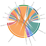
Figure 4. Sankey diagram showing the distribution of Listeria monocytogenes CCs across major food categories in study of clustering and source association of clinical and nonclinical L. monocytogenesisolates,...
Some CCs were significantly associated with certain food categories (Table 2; Figure 4), including raw or processed forms of the same type (e.g., raw or processed dairy). For example, CC9 was significantly associated with processed meat, CC5 with raw meat, and CC121 with processed fish. Other CCs were not statistically associated with certain food categories after correction for multiple testing (false discovery rate–adjusted p value>0.05) but tended to be more commonly found in a specific category. For example, CC6 was ≈11 times more likely to be isolated from processed dairy and 2 times more likely to be isolated from raw dairy compared with all other food categories (Appendix Table 1).
We also performed further comparison for CCs with >10 food isolates (Appendix Table 2) to assess whether those CCs were specifically associated with the processed or raw forms of those key food categories (i.e., meat, fish, dairy). CC9 was more frequently observed among processed meat isolates and CC121 was more frequently observed among processed fish isolates than in raw meat (odds ratio [OR] 15.74 [95% CI 0.90–276.54]) and raw fish (OR 1.40 [95% CI 0.31–6.40]) isolates; however, those associations were not statistically significant after correction for multiple testing. Conversely, CC5 isolates were significantly overrepresented among raw meat compared with processed meat isolates (OR 20 [95% CI 4.17–100]), even after false discovery rate correction.
Virulence and Sanitizer-Tolerance Genes
We screened clinical and nonclinical isolates for virulence and sanitizer tolerance genes (Appendix Figures 1, 2). The Listeria pathogenicity island (LIPI) 1, inlA, and inlB were nearly ubiquitous across both clinical and nonclinical isolates (>98%), whereas LIPI-3 and LIPI-4 showed higher prevalence among clinical isolates (43.64% for LIPI-3 and 19.48% for LIPI-4). We detected inlA premature stop codons (PMSCs) in 32 (3.18%) clinical and 217 (19.92%) nonclinical isolates; frequency was high among nonclinical-associated CCs such as CC9 (91%), CC199 (93%), and CC321 (97.50%). Among clinical isolates with inlA PMSC, 17 (53.12%) carried PMSC type 3 (truncated to 699 aa vs. the full-length 800 aa) and only 2 (6.25%) carried PMSC type 4 (8 aa). Among nonclinical isolates with inlA PMSC, PMSCs type 4 (79 [36.40%]) and type 3 (78 [35.94%]) predominated. PMSC type 4 was rarer among clinical isolates (n = 3 [0.30% of clinical isolates]) than in nonclinical isolates (n = 79 [7.25% of nonclinical isolates]). The remaining PMSCs spanned 14 other PMSC types, resulting in InlA proteins of varying lengths (Table 3). Sanitizer tolerance genes (bcrABC, Tn6188_qac, or LGI1_LM5578_1862) were detected in 354 nonclinical isolates (32.51% of nonclinical isolates; OR 5.81 [95% CI 4.44–7.67]). We noted significant overrepresentation of inlA PMSCs among processed meat isolates (Table 4). In contrast, inlA PMSCs were rare or absent among dairy product isolates (0 of 47 raw dairy isolates; 1 of 21 processed dairy isolates) (Table 4). Sanitizer tolerance genes were significantly overrepresented among isolates from processed fish (OR 3.00 [95% CI 1.74–5.16]) and underrepresented among raw dairy isolates (OR 0.02 [95% CI 0–0.30]) (Appendix Table 3).
Influence of Food Category on Time between Isolate Collection and Detection of Genetically Related Clinical Isolate
We used a Cox proportional hazards model to evaluate the time between food isolate collection and subsequent detection of a genetically related clinical isolate (<50 SNPs) for food categories with >10 isolates (Appendix). Overall, we identified 205 food–clinical isolate pairs; 43 distinct food isolates (13 processed meat, 12 processed fish, 6 raw dairy, 4 processed dairy, 4 produce, 3 salad, and 1 raw meat) were linked with 107 distinct clinical isolates, 1 of which was linked with multiple food sources (processed meat and processed fish) at the same SNP distance. Processed meat (n = 126 pairs) and processed fish (n = 23 pairs) isolates had the highest number of links to clinical isolates (Table 5). The 205 food–clinical pairs were observed in 24 distinct SNP clusters and represented 17 distinct CCs (Table 5). Among the 43 food isolates, 10 of 13 processed meat isolates and 9 of 12 processed fish isolates had inlA PMSC. Out of 205 food–clinical isolate pairs, only 48 (23%) pairs with 16 distinct clinical isolates were involved in 4 different formal outbreak investigations. Among those, only 2 outbreak investigations had implicated food sources (processed meat in both outbreaks) for 13 clinical isolates; for those 2 outbreaks, the implicated food sources were consistent with the food category (i.e., processed meat) identified in our analysis.
Figure 5
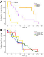
Figure 5. Kaplan-Meier survival curves for the time between food isolate collection and subsequent detection of a genetically related clinical Listeria monocytogenesisolate (single-nucleotide polymorphisms <50) by food category in study...
Food category, but not the presence of inlA PMSC, was a significant predictor of the time between food isolate collection and subsequent detection of a genetically related clinical isolate. In particular, produce isolates exhibited the shortest time for subsequent detection of a genetically related clinical isolate. For example, the hazard ratio for produce compared with processed dairy was 9.72 (95% CI 2.46–38.39), indicating a shorter time between produce isolate collection and subsequent detection of a genetically related clinical isolate than for processed dairy isolates (Appendix Table 4). Survival curves revealed that 75% of produce isolates were detected as genetically related with subsequently detected clinical isolates within 2.2 years; in contrast, some raw meat isolate pairs took up to 17 years to be genetically linked with a clinical isolate (Figure 5, panel A).
Our analysis of L. monocytogenes isolates in NY during 2000–2021 reveals clear differences in CC distributions between clinical and nonclinical isolates. Six CCs were significantly overrepresented among clinical isolates. Of those, 2 CCs (CC1 and CC4) have previously been identified as hypervirulent CCs on the basis of experimental evidence showing that they exhibit enhanced gut colonization, intestinal invasion, and infection of internal organs in a humanized mouse model (4). CC1, a well-established hypervirulent CC associated with invasive listeriosis (14,15), was the most frequently isolated CC among clinical isolates in our study, similar to a previous analysis of NY clinical isolates from 2000–2020 (16). The other CCs significantly overrepresented among clinical isolates in our study (i.e., CC217, CC388, CC389, and CC554) have not been phenotypically classified in terms of their virulence potential but might represent candidates for additional hypervirulent clones, as supported by studies suggesting that the virulence of L. monocytogenes CCs is positively associated with their epidemiologic association with human clinical cases (3). CC6 was the second most frequently isolated CC among clinical isolates but was not among the CCs significantly overrepresented among clinical isolates. That CC has previously been classified as hypervirulent and has been implicated in multiple large listeriosis outbreaks, including outbreaks linked to turkey deli meats, hot dogs, and French-style cheese (17). Previous studies in France and the Netherlands also identified CC1, CC2, CC4, and CC6 as frequently isolated CCs among clinical isolates (3,18). Overall, our data support that certain CCs are more likely to cause human disease.
We also identified 6 CCs (CC5, CC9, CC121, CC199, CC288, and CC321) that were significantly overrepresented among nonclinical isolates. CC9, CC121, CC199, and CC321 were the CCs with the highest proportion of isolates carrying inlA PMSC, a frequent hallmark of hypovirulent L. monocytogenes strains (13,19,20). CC9 and CC121 have previously been described as hypovirulent and environmentally adapted (4); those CCs were also reported to show increased tolerance to benzalkonium chloride, a commonly used sanitizer (4). Two CCs (CC5 and CC288) were overrepresented among nonclinical isolates but exhibited low frequencies of inlA PMSCs. Although CC5 was overrepresented among nonclinical isolates, it was the third most common CC among clinical isolates, suggesting that CC5 isolates are unlikely to be hypovirulent. The observed frequency of CCs among nonclinical isolates differs from observations in Europe. For example, CC121 was the most isolated CC among food and food processing environment–related isolates in France and Norway (3,22), as was CC9 in the Netherlands (18); CC121 and CC9 were the ninth and fifth most common CCs among nonclinical isolates characterized in this study. In contrast, CC5 was the most common CC among nonclinical isolates, but that CC was infrequently found among clinical and nonclinical isolates in France and the Netherlands (3,18). That discrepancy suggests a geographic difference in the distribution of this CC, which could be driven by possible differences in production systems, food preferences, or regional dynamics in L. monocytogenes transmission between NY and those countries in Europe. Despite likely regional differences in prevalence of different CCs, our data further support frequent global occurrence of certain clinically associated CCs (i.e., CC1 and CC4), whereas the occurrence of nonclinically associated CCs seems to show larger regional differences.
Overall, we found 7 statistically significant associations between CCs and food categories. For example, CC9 was significantly overrepresented among processed meat isolates and CC121 was significantly overrepresented among processed fish isolates. Those findings are consistent with previous reports indicating frequent recovery of those CCs from ready-to-eat meat and fish products and infrequent occurrence in dairy products (4,22–24). In addition, CC5 showed overrepresentation among raw meat isolates compared with processed meat isolates. That finding might suggest that different CCs could be introduced at distinct points along the food production chain; some CCs might be more closely linked to primary production or raw materials and others could be more likely to persist in food processing facilities. For the other 5 CCs associated with specific food categories, associations were based on smaller numbers (<10 isolates for the associated food category) and represented associations that had not been previously reported. Thus, those associations are possibly driven by specific sources (rather than general associations with a product type). Overall, our findings support that some CCs might be associated with certain food categories or with raw versus processed foods in a given category, which highlights opportunities to use WGS data to improve outbreak investigations and enable more rapid identification of a likely food source. However, although some CCs showed overrepresentation within specific food categories (e.g., CC9 with processed meat), others (e.g., CC321) were observed across multiple food sources with no overrepresentation in specific food categories. That finding suggests that certain CCs might not be specific to a single reservoir, and their presence should be interpreted cautiously in the context of source attribution.
Overall, inlA PMSCs were significantly overrepresented among isolates from processed meat and overrepresented, but they were not significantly overrepresented among processed fish isolates. Although our findings are consistent with previous studies that have reported frequent occurrence of inlA PMSC in isolates from processing plant environments and ready-to-eat foods (13,19,21,25), our analysis further highlights significant differences between processed and raw food isolates in the frequency of inlA PMSCs. Of note, a previous study in France also reported that hypovirulent CCs were associated with meat products in general (without clear indications on raw vs. processed sources) and proposed that hypovirulent CCs might be adapted to the processing environment, whereas hypervirulent isolates might be adapted to hosts (4). In contrast, we found that intact inlA were overrepresented among dairy isolates, which is also consistent with previous studies, such as a study in France that reported hypervirulent CCs to be strongly associated with dairy products (13). Finally, we found that intact inlA was borderline overrepresented among produce isolates (14 of 15 produce isolates showed intact inlA). That pattern is consistent with produce- and mushroom-focused surveys in the United States (5,26), United Kingdom (27), and China (28), which consistently reported low frequencies of inlA PMSCs in produce-associated isolates (5). In addition, we also found that sanitizer tolerance genes were significantly overrepresented in processed fish isolates and underrepresented in raw dairy isolates, consistent with a previous study that sanitizer tolerance genes were frequent among processed fish isolates (5). Overall, our findings suggest that food categories might differ in their likelihood of carrying virulence-attenuated L. monocytogenes and indicate that using WGS data to identify genetic markers (e.g., inlA PMSCs, sanitizer tolerance genes) could help with source attribution of raw versus finished products for some specific food categories.
We hypothesized that food category and presence of inlA PMSC were associated with the time between a food isolate collection and subsequent detection of a genetically related clinical isolate. Of note, we found that food category, but not the presence of inlA PMSC, was a significant predictor of that time; differences in time interval were observed across food categories. Specifically, isolates from produce had a shorter time for subsequent detection of a genetically related clinical isolate than did isolates from all other food categories.
Several factors likely contribute to the early detection of clinical isolates that are genetically related to produce isolates. Fresh produce typically has a shorter shelf life than other ready-to-eat products (e.g., cheese), possibly creating a narrow window in which contamination can directly lead to human exposure and illness. Contaminated produce typically would be consumed <20 days after processing, whereas cheese could be consumed >6 months after processing. However, the observed difference is unlikely to be explained solely by the short shelf life of fresh produce, because the time scale observed extends well beyond typical produce shelf life. Differences in consumer storage practices across different food categories (e.g., persons would more likely freeze raw meat than fresh produce) might extend the exposure window for certain food categories. In addition, L. monocytogenes contamination might be more frequently driven by repeated reintroduction rather than long-term persistence in packing and fresh-cut facilities (29–31). Persistent L. monocytogenes strains have also been detected less frequently in produce packinghouses (31–33) than in other food-associated facilities, although persistence over multiple seasons in some produce packing houses has been reported (34). Further research is needed to elucidate the persistence of L. monocytogenes in produce environments (29), but our findings suggest somewhat unique L. monocytogenes transmission pathways for produce. Of note, our data also showed that the time between the first food isolate and last subsequent detection of genetically linked clinical isolate is shorter for produce than for other food categories. That finding suggests that produce-linked clusters and outbreaks might often cover relatively short time frames, although long-term multistate outbreaks linked to produce have been reported (35). Even extremely small outbreaks putatively linked to produce might need to be further prioritized for outbreak investigations.
The first limitation of our study is that our dataset only included isolates obtained in NY during 2000–2021. Therefore, the proportion of unclustered and unlinked isolates might be overestimated because some clinical and nonclinical isolates could be clustered or linked with isolates from outside NY. Second, the SNP thresholds used to define clustering and linkage (i.e., <20 SNPs for clinical–clinical isolates and <50 SNPs for clinical–nonclinical isolates) are supported by previous studies as useful initial cut-offs for screening of potential clusters and sources but might need adjustment based on available epidemiologic data, particularly during outbreak investigations. Consequently, genetic relatedness inferred using those thresholds should be interpreted with caution. Third, genetic similarity alone does not establish epidemiologic linkage. Isolates with low SNP distances might reflect persistence in food production environments or widespread dissemination rather than direct transmission events. This study did not incorporate epidemiologic data (e.g., exposure histories), which limits the ability to confirm specific food sources for clinical cases. Similarly, a limitation of the cluster analysis is that some observed intervals between food isolate collection and subsequent detection of a genetically related clinical isolate were long. Those long intervals reduce confidence in interpreting the food as a continuous source of human exposure. Instead, such associations might reflect repeated reintroduction or cross-contamination across different reservoirs or food commodities. That interpretation is further supported by the observation that some SNP clusters included isolates from multiple food categories (Table 5). Therefore, the time intervals should be interpreted as reflecting temporal patterns of genomic relatedness rather than direct evidence of long-term source attribution to a single reservoir or commodity. Fourth, the dataset might be subject to biases, including uneven representation across food categories and time periods. In particular, the dataset includes isolates collected through targeted surveillance and research activities, which might result in overrepresentation of certain food categories or production environments. In addition, although thought to be rare, underdiagnosis or underreporting of listeriosis cases might lead to incomplete capture of clinical isolates. Finally, this study relied solely on SNP-based analyses. Incorporating approaches such as core-genome/whole-genome MLST could further strengthen the robustness of the findings, which highlights the need for more comprehensive integration of clinical and nonclinical isolates into unified databases to enable consistent genomic analyses, clustering, and outbreak investigations. In the future, advances in artificial intelligence and machine learning might further enhance the interpretation of surveillance datasets by identifying potential source attribution that are not easily analyzed through conventional approaches.
Ms. Samut is a PhD student in food science at Cornell University with a research focus on foodborne bacterial pathogens. Her research interests include molecular epidemiology and evolutionary dynamics of Salmonella enterica and Listeria monocytogenes.
Acknowledgments
We thank the staff of the Advanced Genomic Technologies Cluster at the Wadsworth Center where library preparation and sequencing were carried out.
For New York State Department of Agriculture and Markets, the whole-genome sequencing was supported by the Food and Drug Administration (FDA) of the US Department of Health and Human Services (HHS) as part of a financial assistance award (5U19FD007122), with 100% funded by FDA/HHS. The contents are those of the authors and do not necessarily represent the official views of, nor an endorsement, by FDA/HHS or the US government. For New York State Department of Health, this work was supported by New York State, the Centers for Disease Control and Prevention Epidemiology and Laboratory Capacity Grant (cooperative agreement no. NU50CK000423), and the Food and Drug Administration’s LFFM Grant (cooperative agreement no. 1U19FD007089). For Cornell University, this work was supported by an ELC Foodborne Illness Centers of Excellence grant (no. 5NU50CK000516-05-00) awarded by Health Research Inc.
References
- Scallan Walter EJ, Cui Z, Tierney R, Griffin PM, Hoekstra RM, Payne DC, et al. Foodborne illness acquired in the United States—major pathogens, 2019. Emerg Infect Dis. 2025;31:669–77.PubMedGoogle Scholar
- Ferreira V, Wiedmann M, Teixeira P, Stasiewicz MJ. Listeria monocytogenes persistence in food-associated environments: epidemiology, strain characteristics, and implications for public health. J Food Prot. 2014;77:150–70. DOIPubMedGoogle Scholar
- Maury MM, Tsai YH, Charlier C, Touchon M, Chenal-Francisque V, Leclercq A, et al. Uncovering Listeria monocytogenes hypervirulence by harnessing its biodiversity. Nat Genet. 2016;48:308–13. DOIPubMedGoogle Scholar
- Maury MM, Bracq-Dieye H, Huang L, Vales G, Lavina M, Thouvenot P, et al. Hypervirulent Listeria monocytogenes clones’ adaption to mammalian gut accounts for their association with dairy products. Nat Commun. 2019;10:2488. DOIPubMedGoogle Scholar
- Daeschel D, Pettengill JB, Wang Y, Chen Y, Allard M, Snyder AB. Genomic analysis of Listeria monocytogenes from US food processing environments reveals a high prevalence of QAC efflux genes but limited evidence of their contribution to environmental persistence. BMC Genomics. 2022;23:488. DOIPubMedGoogle Scholar
- He Y, Xu T, Li S, Mann DA, Britton B, Oliver HF, et al. Integrative assessment of reduced Listeria monocytogenes susceptibility to benzalkonium chloride in produce processing environments. Appl Environ Microbiol. 2022;88:
e0126922 . DOIPubMedGoogle Scholar - Stout A, Van Stelten-Carlson A, Marquis H, Ballou M, Reilly B, Loneragan GH, et al. Public health impact of foodborne exposure to naturally occurring virulence-attenuated Listeria monocytogenes: inference from mouse and mathematical models. Interface Focus. 2020;10:
20190046 . DOIPubMedGoogle Scholar - Samut H, Mendez-Vallellanes DV, Hoyt H, Wirth SE, Mingle L, Sauders BD, et al. Retrospective analysis of historical Listeria monocytogenes clinical isolates, New York, USA, 2000–2021. Emerg Infect Dis. 2025;31:1883–92. DOIPubMedGoogle Scholar
- Pightling AW, Pettengill JB, Luo Y, Baugher JD, Rand H, Strain E. Interpreting whole-genome sequence analyses of foodborne bacteria for regulatory applications and outbreak investigations. Front Microbiol. 2018;9:1482. DOIPubMedGoogle Scholar
- Jackson KA, Gould LH, Hunter JC, Kucerova Z, Jackson B. Listeriosis outbreaks associated with soft cheeses, United States, 1998–2014. Emerg Infect Dis. 2018;24:1116–8. DOIPubMedGoogle Scholar
- Chen Y, Gonzalez-Escalona N, Hammack TS, Allard MW, Strain EA, Brown EW. Core genome multilocus sequence typing for identification of globally distributed clonal groups and differentiation of outbreak strains of Listeria monocytogenes. Appl Environ Microbiol. 2016;82:6258–72. DOIPubMedGoogle Scholar
- Pettengill JB, Rand H, Wang SS, Kautter D, Pightling A, Wang Y. Transient and resident pathogens: Intra-facility genetic diversity of Listeria monocytogenes and Salmonella from food production environments. PLoS One. 2022;17:
e0268470 .PubMedGoogle Scholar - Van Stelten A, Simpson JM, Ward TJ, Nightingale KK. Revelation by single-nucleotide polymorphism genotyping that mutations leading to a premature stop codon in inlA are common among Listeria monocytogenes isolates from ready-to-eat foods but not human listeriosis cases. Appl Environ Microbiol. 2010;76:2783–90. DOIPubMedGoogle Scholar
- Moura A, Lefrancq N, Wirth T, Leclercq A, Borges V, Gilpin B, et al. Listeria CC1 Study Group. Emergence and global spread of Listeria monocytogenes main clinical clonal complex. Sci Adv. 2021;7:
eabj9805 . DOIPubMedGoogle Scholar - Fotopoulou ET, Jenkins C, Barker CR, Painset A, Didelot X, Simbo A, et al. Genomic epidemiology of the clinically dominant clonal complex 1 in the Listeria monocytogenes population in the UK. Microb Genom. 2024;10:
001155 . DOIPubMedGoogle Scholar - Ikhimiukor OO, Mingle L, Wirth SE, Mendez-Vallellanes DV, Hoyt H, Musser KA, et al. Long-term persistence of diverse clones shapes the transmission landscape of invasive Listeria monocytogenes. Genome Med. 2024;16:109. DOIPubMedGoogle Scholar
- Chen Y, Gonzalez-Escalona N, Hammack TS, Allard MW, Strain EA, Brown EW. Core genome multilocus sequence typing for identification of globally distributed clonal groups and differentiation of outbreak strains of Listeria monocytogenes. Appl Environ Microbiol. 2016;82:6258–72. DOIPubMedGoogle Scholar
- Coipan CE, Friesema IHM, van Hoek AHAM, van den Bosch T, van den Beld M, Kuiling S, et al. New insights into the epidemiology of Listeria monocytogenes—a cross-sectoral retrospective genomic analysis in the Netherlands (2010–2020). Front Microbiol. 2023;14:
1147137 . DOIPubMedGoogle Scholar - Nightingale KK, Windham K, Martin KE, Yeung M, Wiedmann M. Select Listeria monocytogenes subtypes commonly found in foods carry distinct nonsense mutations in inlA, leading to expression of truncated and secreted internalin A, and are associated with a reduced invasion phenotype for human intestinal epithelial cells. Appl Environ Microbiol. 2005;71:8764–72. DOIPubMedGoogle Scholar
- Magagna G, Gori M, Russini V, De Angelis V, Spinelli E, Filipello V, et al. Evaluation of the virulence potential of Listeria monocytogenes through the characterization of the truncated forms of Internalin A. Int J Mol Sci. 2023;24:10141. DOIPubMedGoogle Scholar
- Manuel CS, Van Stelten A, Wiedmann M, Nightingale KK, Orsi RH. Prevalence and distribution of Listeria monocytogenes inlA alleles prone to phase variation and inlA alleles with premature stop codon mutations among human, food, animal, and environmental isolates. Appl Environ Microbiol. 2015;81:8339–45. DOIPubMedGoogle Scholar
- Fagerlund A, Wagner E, Møretrø T, Heir E, Moen B, Rychli K, et al. Pervasive Listeria monocytogenes is common in the Norwegian food system and is associated with increased prevalence of stress survival and resistance determinants. Appl Environ Microbiol. 2022;88:
e0086122 . DOIPubMedGoogle Scholar - Wu J, NicAogáin K, McAuliffe O, Jordan K, O’Byrne C. NicAogáin K, McAuliffe O, Jordan K, O’Byrne C. NicAogáin K, McAuliffe O, Jordan K, O’Byrne C. Phylogenetic and phenotypic analyses of a collection of food and clinical Listeria monocytogenes isolates reveal loss of function of sigma B from several clonal complexes. Appl Environ Microbiol. 2022;88:
e0005122 . DOIPubMedGoogle Scholar - Pérez-Baltar A, Pérez-Boto D, Medina M, Montiel R. Genomic diversity and characterization of Listeria monocytogenes from dry-cured ham processing plants. Food Microbiol. 2021;99:
103779 . DOIPubMedGoogle Scholar - Wagner E, Fagerlund A, Thalguter S, Jensen MR, Heir E, Møretrø T, et al. Deciphering the virulence potential of Listeria monocytogenes in the Norwegian meat and salmon processing industry by combining whole genome sequencing and in vitro data. Int J Food Microbiol. 2022;383:
109962 . DOIPubMedGoogle Scholar - Amarasekara NR, Swamy AS, Paudel SK, Jiang W, Li K, Shen C, et al. Hypervirulent clonal complex (CC) of Listeria monocytogenes in fresh produce from urban communities. Front Microbiol. 2024;15:
1307610 . DOIPubMedGoogle Scholar - Smith A, Hearn J, Taylor C, Wheelhouse N, Kaczmarek M, Moorhouse E, et al. Listeria monocytogenes isolates from ready to eat plant produce are diverse and have virulence potential. Int J Food Microbiol. 2019;299:23–32. DOIPubMedGoogle Scholar
- Chen M, Cheng J, Wu Q, Zhang J, Chen Y, Zeng H, et al. Prevalence, potential virulence, and genetic diversity of Listeria monocytogenes isolates from edible mushrooms in Chinese markets. Front Microbiol. 2018;9:1711. DOIPubMedGoogle Scholar
- Belias A, Sullivan G, Wiedmann M, Ivanek R. Factors that contribute to persistent Listeria in food processing facilities and relevant interventions: a rapid review. Food Control. 2022;133:133. DOIGoogle Scholar
- Sullivan G, Orsi RH, Estrada E, Strawn L, Wiedmann M. Whole-genome sequencing-based characterization of Listeria isolates from produce packinghouses and fresh-cut facilities suggests both persistence and reintroduction of fully virulent L. monocytogenes. Appl Environ Microbiol. 2022;88:
e0117722 . DOIPubMedGoogle Scholar - Estrada EM, Hamilton AM, Sullivan GB, Wiedmann M, Critzer FJ, Strawn LK. Prevalence, persistence, and diversity of Listeria monocytogenes and Listeria species in produce packinghouses in three U.S. states. J Food Prot. 2020;83:277–86. DOIPubMedGoogle Scholar
- Murugesan L, Kucerova Z, Knabel SJ, LaBorde LF. Predominance and distribution of a persistent Listeria monocytogenes clone in a commercial fresh mushroom processing environment. J Food Prot. 2015;78:1988–98. DOIPubMedGoogle Scholar
- Chen Y, Simonetti T, Peter K, Jin Q, Brown E, LaBorde LF, et al. Genetic diversity of Listeria monocytogenes isolated from three commercial tree fruit packinghouses and evidence of persistent and transient contamination. Front Microbiol. 2022;12:
756688 . DOIPubMedGoogle Scholar - Simonetti T, Peter K, Chen Y, Jin Q, Zhang G, LaBorde LF, et al. Prevalence and distribution of Listeria monocytogenes in three commercial tree fruit packinghouses. Front Microbiol. 2021;12:
652708 . DOIPubMedGoogle Scholar - Palacios A, Vasser M, Madad A, Ivory S, Edwards L, Donovan D, et al. Two concurrent outbreaks of Listeria monocytogenes infections linked to packaged salads, United States, 2014–2022. Emerg Infect Dis. 2025;31:2216–24. DOIPubMedGoogle Scholar
Figures
Tables
Cite This ArticleOriginal Publication Date: August 17, 2026
1Preliminary results from this study were presented at the 2025 International Association for Food Protection Conference; July 27–30, 2025; Cleveland, Ohio, USA.
Table of Contents – Volume 32, Number 9—September 2026
| EID Search Options |
|---|
 Advanced Article Search – Search articles by author and/or keyword.
|
 Articles by Country Search – Search articles by the topic country.
|
 Article Type Search – Search articles by article type and issue.
|
Please use the form below to submit correspondence to the authors or contact them at the following address:
Renato H. Orsi, Department of Food Science, Cornell University, 326 Stocking Hall, Ithaca, NY 14853, USA
Top