Volume 32, Number 8—August 2026
Research
Virulence of Burkholderia pseudomallei Strains from Western Hemisphere and Africa in Mice


Cite This Article
Citation for Media
Abstract
Melioidosis, caused by Burkholderia pseudomallei, is an emerging disease in the United States and was declared endemic to the Gulf Coast region in 2022. Melioidosis has been sporadically reported in the American continents, the Caribbean, and more recently in Africa. We conducted lethal dose analyses in BALB/c and C57BL/6 mice exposed to small particle aerosols of B. pseudomallei strains from the Western Hemisphere and Africa. Those isolates exhibit a variety of virulence patterns, including rapidly-lethal disease and delayed onset of fatal disease. We found that the isolates we tested grew similarly in culture but displayed major differences in biofilm formation. Our data contribute to the growing knowledge of geographically distinct B. pseudomallei isolates and aid in ensuring that medical countermeasures in development are effective against a diverse collection of bacterial strains.
Burkholderia pseudomallei, a gram-negative facultative intracellular bacterium, causes the disease melioidosis and is critical to both public health and biodefense research communities (1). Historically, melioidosis is a public health concern in Southeast Asia and northern Australia; however, the global range of melioidosis is much larger than previously described (2–4). Melioidosis is highly underreported in many areas around the world, including India, Africa, the Caribbean, and the Americas (5–16). Underreporting can be attributed to extremely diverse clinical manifestations, scarcity of appropriate diagnostic tools, and a general lack of knowledge of B. pseudomallei by medical staff in endemic regions outside of the historically prevalent areas. However, frequent case reports detailing new clinical examples of melioidosis underscore the importance of this disease throughout the world, particularly in developing nations. B. pseudomallei is also considered an emerging threat in the United States because of several evolving factors: endemicity in the Gulf Coast region and the Caribbean territories of Puerto Rico and US Virgin Islands (7,11,17); documented fatal cases resulting from unintentional importation of the bacterium to the United States by an aromatherapy spray produced in India (18); evidence of locally acquired melioidosis in Texas (12); and recent infections in Georgia after a severe weather event (19).
We examined 10 phylogeographically diverse strains of B. pseudomallei from the Western Hemisphere and 3 strains originating from Ghana in Africa. We performed in vitro growth analyses and biofilm assays of those isolates and calculated 50% lethal dose (LD50) estimations in both the BALB/c and C57BL/6 mouse models of inhalational melioidosis. BALB/c mice are considered an acute model (or less resistant to B. pseudomallei) and are the model of choice for therapeutic medical countermeasure development (20). In contrast, C57BL/6 mice are considered a more chronic infection model and are the generally accepted model for vaccine development (21–25).
Bacterial In Vitro Growth Conditions and Assessment
We grew B. pseudomallei strains in 4% glycerol/1% tryp¬tone/0.5% NaCl broth for 16 hours at 37°C, shaking at 200 rpm for in vivo testing. We grew B. pseudomallei strains in Luria broth with 4% glycerol (LBG) to assess in vitro growth kinet¬ics. We resuspended the strains from an overnight broth culture at 37°C to a 600 nm optical density (OD600) of 0.2 in LBG.e resuspended the strains from an overnight broth culture at 37°C to a 600 nm optical density (OD600) of 0.2 in LBG. We diluted suspensions 1:10 into a 96-well microtiter plate and cultured in a Spark microplate reader (Tecan Group, https://www.tecan.com) with shaking at 37°C for ≈24 hours. We measured the OD600 in 15-minute intervals. We determined the actual density by subtracting the value of the respective medium-only control from the measured OD600.
Biofilm Assays
We used crystal violet staining to quantify biofilm production. We adjusted overnight B. pseudomallei LBG broth cultures to an OD600 of ≈0.2, diluted 1:10 into LBG, and then incubated at 37°C for 24 hours. Prior to staining, we measured the OD600 of the bacterial suspension, then aspirated the plates, washed them 3 times with phosphate buffered saline to remove planktonic cells, and fixed them with 100% ethanol for 30 minutes at room temperature (26). To stain the biofilm, we added 0.1% crystal violet (wt/vol) to each well for 15 min and washed 3 times with phosphate buffered saline; we then solubilized the remaining stain in 33% acetic acid. To quantify staining, we measured the OD600 as an indicator of biofilm formation. When necessary, we diluted the samples in 33% acetic acid to ensure that readings were within linear range. We collected >3 technical replicates for each of 4 individual repeated experiments and averaged the values for each experiment. We used control B. pseudomallei strains K96243 and ATS2021 as comparators to test strains with different biofilm profiles.
Mouse Median Lethal Dose Estimations
We exposed 7–9-week-old female BALB/c or C57BL/6 mice (n = 10/group) to 5 different doses of aerosolized strains of B. pseudomallei. Starting-dose concentrations for each target dose in the lethal dose (LD50) estimate ranged from ≈104 to 109 colony forming units (CFU)/mL. We generated small particle aerosols (1–3 μm) by using the Biaera system (Biaera, https://www.biaera.com). We then calculated the estimated inhaled doses by using Guyton’s formula based on the CFU/mL determination of an all-glass impinger collection medium (27–29). We monitored the mice 1 time/day for animal husbandry purposes and >1 time/day for clinical scoring. Scores of 0–2 represented normal mice; scores of 3–7 indicated major clinical manifestations (e.g., less peer interaction, reduced grooming, labored breathing), and those mice warranted multiple clinical assessments per day; and final scores of >8 indicated severe clinical manifestations, and those mice were euthanized immediately. We euthanized mice in accordance with early intervention criteria. At the end of the study, we euthanized the surviving mice. We enumerated residual bacteria in lungs, spleens, and brains in select dose-groups to determine if there were any novel bacterial dissemination patterns or evidence of unusual chronicity associated with any of the previously uncharacterized strains of B. pseudomallei.
This research was conducted under an Institutional Animal Care and Use Committee approved protocol in compliance with the Animal Welfare Act, Public Health Service Policy on Humane Care and Use of Laboratory Animals, and other federal statutes and regulations relating to animals and experiments involving animals. The facility where this research was conducted is accredited by Association for Assessment and Accreditation of Laboratory Animal Care International and adheres to the principles stated in The Guide for the Care and Use of Laboratory Animals, National Research Council, 2011.
Statistics
We used probit analysis to estimate the LD50 by using log10 dose as the predictor. We tested the effect of B. pseudomallei strain and mouse on LD50 by using a χ2 test on the basis of the probit model and under the assumption of a common effect of log10 dose. We estimated median time-to-death (TTD) or euthanasia and accompanying confidence limits by using Kaplan-Meier survival methods. We performed comparisons between B. pseudomallei strains by using Wald tests of least squared mean differences on the basis of a log-normal accelerated failure time model. We calculated the Pearson and Spearman rank-order correlation analysis of the geometric mean of biofilm versus log of LD50. We compared pairwise treatment groups by using linear mixed effects model for biofilm comparisons. We adjusted the multiplicity by using the Tukey method. We implemented analysis by using SAS version 9.4 (SAS Institute Inc., https://www.sas.com).
In Vitro Growth and Biofilm Analyses
Figure 1
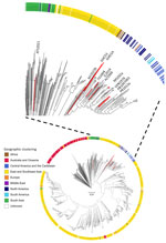
Figure 1. Maximum-likelihood core-genome phylogenetic tree of all Burkholderia pseudomallei genomes (n = 1,956) from the National Center for Biotechnology Information RefSeq (https://www.ncbi.nlm.nih.gov/refseq) database used in study of ...
Figure 2
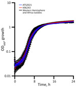
Figure 2. Western Hemisphere and Africa test panel isolates in a study of Burkholderia pseudomallei virulence in mouse models showing no differences in planktonic growth. B. pseudomalleiwas grown...
Figure 3
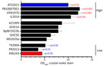
Figure 3. Observations of biofilm formation in a study of Burkholderia pseudomallei virulence in mouse models. Burkholderia pseudomalleiisolates were inoculated into Luria broth with 4% glycerol and incubated...
We assembled a new panel of B. pseudomallei strains (Table 1) that are phylogeographically diverse. Our results underscore the phylogenetic diversity and geographic origin of the strains included in this study (Figure 1). We assessed the in vitro growth rates in LBG and identified near-identical growth rates for each isolate (Figure 2). In our evaluation of biofilm production, the different geographic isolates exhibited highly different biofilm production (Figure 3). We included 2 well-characterized strains, ATS2021 and K96243, for comparison. The ATS2021 strain was demonstrated as a hyper-biofilm producer (26), and the K96243 strain is one of the most used strains in melioidosis research.
In Vivo Median LD50 Estimations
Figure 4
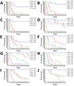
Figure 4. Survival rates for aerosolized challenge of Western Hemisphere and Africa Burkholderia pseudomallei strain virulence in BALB/c mouse models. We challenged BALB/c mice with 5 different doses of aerosolized ...
Figure 5
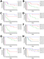
Figure 5. Survival rates for aerosolized challenge of Western Hemisphere and Africa Burkholderia pseudomallei strain virulence in C57BL/6 mouse models. C57BL/6 mice were challenged with 5 different doses of aerosolized ...
We calculated LD50 values for each isolate in both BALB/c and C57BL/6 mice on day 21 and day 60 to help gauge the infection lethality during the first several weeks postexposure (acute phase) compared with the lethality observed throughout the entire 60-day study (Table 2; Figures 4, 5; Appendix 1 Tables 1, 2). The first pattern we observed from the dataset was that the 3 B. pseudomallei strains from Ghana (GHD1A, BpNY2023b, and GHC5E) displayed low LD50 estimates with rapid disease (day 21, <5 CFU/mL for BALB/c mice, 160–360 CFU/mL for C57BL/6 mice) and were among the most acutely lethal strains in the mouse model of inhalational melioidosis (day 60, 1–3 CFU/mL for BALB/c mice, 79–113 CFU/mL for C57BL/6 mice). The B. pseudomallei strain from Costa Rica (PB10007001) was approximately equally virulent when LD50 was compared with strains from Ghana (p>0.05). Second, the B. pseudomallei strains tested from Venezuela (VEN1976), Puerto Rico (PR2013a), and Mississippi (MS2020a) appeared less virulent during the first few weeks but continued to cause death through the 60-day study period, ultimately demonstrating major virulence by the study end. Last, relative to the other B. pseudomallei strains in this panel, the strains from Texas (TX2004), El Salvador (AZ1999), and Mexico (IL2014) exhibited slowly progressive virulence or displayed persistence-associated phenotypes at both the day 21 and day 60 postexposure to aerosolized bacteria. Of note, unlike the other B. pseudomallei strains tested, the strain from Mexico (IL2014) did not exhibit expected differential virulence in the 2 mouse strains. Instead, the LD50 estimates for IL2014 were virtually identical on day 21 (ratio of LD50 of 0.9; p = 0.87); although that ratio did increase to 6.3 (p = 0.0045) by the end of the 60-day study, the day 21 data are noteworthy (Table 2; Appendix 1 Table 1). Furthermore, analysis of TTD or euthanasia among all dose-groups identified that those 3 B. pseudomallei strains also had protracted TTD estimates. For example, at the completion of the study, the TX2004 strain had a median TTD of 32.5 (10,33) days in BALB/c mice for the highest dose evaluated, whereas all other strains had a median TTD of <5 at the highest dose tested. Similarly, strains AZ1999, IL2014, MS2020a, and VEN1976 offered additional examples of protracted TTD values (Appendix 1 Table 2).
Determination of Residual Bacteria
At the end of the study (day 60), to identify potential unique bacterial dissemination patterns or evidence of pronounced chronicity in any of the uncharacterized B. pseudomallei strains, we evaluated the bacterial content of the lungs, spleens, and brains from surviving mice in select dose-groups. Our results (Tables 3, 4) were not entirely unexpected on the basis of previous B. pseudomallei panels (28,34), but we identified several trends of note. In the BALB/c mice, only the B. pseudomallei isolates that caused a slower progressing disease (AZ1999, IL2014, and TX2004) resulted in residual bacterial counts at the end of the 60-day study. B. pseudomallei strain PR2013a was an exception with the lowest dose examined (Table 3). As expected, the C57BL/6 mouse model had many more mice harboring bacteria at the end of the 60-day study (Table 4). Nearly every strain and dose we sampled at the end of study had evidence of bacteria in the brains of C57BL/6 mice, and more of the brains remained colonized compared with spleens. Although there are clear anatomic differences between mice and humans that could lead to artifactual bacterial pathogenesis, this prevalence of brain colonization should be considered a critical aspect of this model that could offer a mechanism to evaluate the effectiveness of medical countermeasures against neurologic melioidosis.
Genome Analyses
Figure 6
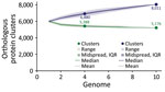
Figure 6. Pangenome analysis of Burkholderia pseudomallei strains from the Western Hemisphere and Africa in mouse models. We used panaroo version 1.5.2 (https://github.com/gtonkinhill/panaroo) for analysis of all 10...
We conducted pangenome analysis of all 10 isolate genomes included in this study to identify gene candidates that could potentially account for the increased virulence associated with the 3 strains of African origin (GHD1A, GHC5E, and BpNY2023b). Whole-genome sequence accession numbers are included (Table 1). The core genome was determined to consist of 5,176 genes, whereas the pangenome was determined to consist of 8,011 total genes (Figure 6). We then further investigated the pangenome and identified genes unique to GHD1A, GHC5E, and BpNY2023b, finding 2 predicted hypothetical protein-encoding genes. Further motif queries of those 2 predicted protein-encoding genes suggest one 753-bp gene makes a transcriptional regulator and the other appears to be a truncated (255-bp) kumamolysin protease.
Our results are a comprehensive evaluation of virulence assessed in mouse models of inhalational melioidosis for strains of B. pseudomallei originating in the Western Hemisphere and Africa. The B. pseudomallei strains we evaluated offer a spectrum of virulence patterns; the Ghana strains uniformly produced acutely lethal disease despite unique genotypes and years between isolation (30,35). Our genomic analyses did not identify clear functional differences at the gene level that definitively distinguish among those strains of B. pseudomallei.
Of note, this study evaluated virulence in mice after exposure to small particle aerosols; however, it does not address virulence of the novel B. pseudomallei strains after ingestion or inoculation via parenteral routes. We did not observe any in vitro growth differences between the strains in the new test panel and the growth kinetics are remarkably similar to previously reported for strains originating in Thailand and northern Australia (34). Previously, when using a panel of B. pseudomallei strains originating from Australia and Thailand, we described a significant inverse correlation with biofilm production and virulence when assessed in mouse models of inhalational melioidosis (26). However, with this current panel of strains, we did not observe this correlation following the same challenge method. More work is required to ascertain if the correlation with inhalation virulence of B. pseudomallei is biologically relevant or if it is only associated with strains from Thailand or Northern Australia.
The role of the B. pseudomallei biofilm has been debated (26,36). However, typically biofilm is considered a critical virulence factor because it enables the bacterial community to withstand the hostile environment within a host and resist antimicrobial drugs (37). For instance, in vitro biofilm formation was found to be higher for isolates obtained from patients with relapsing melioidosis compared with isolates of patients without relapse (38). Further, biofilm forming capacity was implicated in antimicrobial drug susceptibility, although upregulation of biofilm-related genes might play a larger role in antimicrobial drug resistance than the biofilm matrix in B. pseudomallei (33,39).
This analysis of B. pseudomallei strain virulence observed in strains from the Western hemisphere and Africa resulted in similar observations to those reported when examining strains from Thailand and northern Australia (28,34). Some strains are rapidly fatal in the mouse model, and the LD50 estimate remains similar throughout the course of the 60-day study (i.e., GHD1A, BpNY2023b, PB10007001, and GHC5). In contrast, other strains cause disease but require additional time to result in lethality (i.e., AZ1999, VEN1976, PR2013a, and MS2020a); 1 strain (IL2014) had very similar LD50 estimates at day 21 in both BALB/c and C57BL/6 mice. More work is required to understand the similar virulence in both mouse models seen with strain IL2014, but those findings mirror what we have previously described regarding the virulence of strain 1106a from Thailand (28,40). Those unique virulence patterns will be necessary to better characterize the mouse models of inhalational melioidosis and host immunity to this disease.
In our geographically diverse panel, we did not identify patterns that suggested differential virulence associated with isolate origin (i.e., clinical vs. environmental isolate), but we note that these differences have been documented (41,42). For example, when we compared the virulence of the 3 isolates from Ghana (2 environmental isolates with different genomic signatures and 1 human clinical isolate from 2023), we calculated similar median lethal dose estimates determined by the mouse in vivo models (p>0.22 in all cases). B. pseudomallei genomic analyses are complex, and additional analyses will be required to further identify genomic features that could lead to the classification of geographic isolates on the basis of virulence (40,43).
Virulence is difficult to define in B. pseudomallei. Complex acute clinical signs coupled with poorly characterized chronic or latent stages make melioidosis challenging to diagnose in humans and difficult to model in laboratory animals. We observed mice with inhalational melioidosis for 60 days, and in our estimation, that prolonged duration still might not be adequate to capture the unique disease phenotypes exhibited in those animals. Several examples of human case studies question the description of B. pseudomallei infection staying completely latent or dormant for decades, as opposed to the established pattern of relapsing or remitting chronic infection (12,44–46). Of interest, several of those examples include residents of the Gulf Coast region of the United States. Recently, an investigation estimated that activation from a truly asymptomatic infection represented <1.7%–2.9% of cases (47). Improved data collection and analyses, the reevaluation of seminal melioidosis case reports, and the growing dataset of B. pseudomallei in parts of the world outside of northern Australia and Southeast Asia further supports a much larger effect of melioidosis globally than previously estimated and underscores the complexities of melioidosis modeling or virulence assessment.
Our intent is not to oversimplify virulence attributes associated with B. pseudomallei isolates solely by rank ordering median lethal doses in mouse models. Understanding the virulence of this organism will require in-depth and comprehensive studies designed to examine macrophage infection assays, intracellular survival or replication assessments, resistance to host killing, and serial sampling natural history studies in appropriate animal models of disease. When taken together, those data would then offer a more complete view of virulence, particularly in context of other critical parameters that include but are not limited to host-inflammatory response, tissue pathology, bacterial burden or dissemination, and tissue tropisms that might be associated with isolates (48,49). Nevertheless, this study is a comprehensive attempt to begin to understand the virulence associated with Western Hemisphere and African isolates of B. pseudomallei. Of interest is the residual levels of bacteria in the brains of mice (Tables 3, 4). Every strain examined could be found in the brains of the C57BL/6 mice exposed to aerosolized B. pseudomallei (Table 4). Those data reinforce that the C57BL/6 model is less acute than the BALB/c model, likely enabling differential bacterial dissemination and replication in tissues such as the brain. Neurologic impacts are a devastating aspect of inhalational melioidosis; a better understanding of such neuropathogenesis and model refinement could have massive implications for modeling neurologic melioidosis (50; Appendix 2 reference 51).
Although already a regulated biologic select agent because of biodefense concerns, B. pseudomallei is considered an emerging threat in the United States after being declared endemic to the Gulf Coast region (11). Historically, melioidosis has been attributed to travel outside of the continental United States, previously residing in an endemic part of the world, or rare exposure to imported products or a single case associated with an aquarium (32; Appendix 2 references 52–54). The recent lethal cases of melioidosis associated with the unintentional importation of B. pseudomallei in aromatherapy spray manufactured in India were other examples of domestic melioidosis from an unexpected source (18,26). As we continue to understand the risk associated with understudied Western Hemisphere and African strains, it is imperative to perform additional studies. Those data are necessary for medical modeling purposes, risk assessments, and to further educate medical providers in areas of the world where B. pseudomallei is not an expected pathogen (Appendix 2 reference 55).
In conclusion, melioidosis is a challenging disease to study because of its tremendously diverse clinical manifestations in both human patients and laboratory animal models. For the best possible patient outcome, it is vital that this disease is accurately diagnosed and appropriate antimicrobial drug treatments are provided (Appendix 2 references 56,57). In addition to being an emerging threat globally, melioidosis also offers an excellent example of the necessity of continuing research into infectious diseases and challenging accepted dogma as new data become available. The concept that pathogens can be studied and treated with effective novel or repurposed medical countermeasures but research on the organism abandoned because of the treatment successes is counterproductive to both biodefense and public health fields of research. We must continue to monitor biothreats, reexamine risk assessments and disease modeling efforts, and classify or reclassify emerging and reemerging threats to be optimally prepared for future adversarial use, outbreaks, or pandemics.
Mr. Klimko is a scientist at the US Army Medical Research Institute of Infectious Diseases and began his career as an active-duty military service member. His research interests include medical countermeasure development for bacterial biothreat agents and animal modeling under high-containment conditions.
Acknowledgment
This study was funded by the US Defense Threat Reduction Agency (project nos. CB11520 and CB11395).
References
- Meumann EM, Limmathurotsakul D, Dunachie SJ, Wiersinga WJ, Currie BJ. Burkholderia pseudomallei and melioidosis. Nat Rev Microbiol. 2024;22:155–69. DOIPubMedGoogle Scholar
- Limmathurotsakul D, Golding N, Dance DA, Messina JP, Pigott DM, Moyes CL, et al. Predicted global distribution of Burkholderia pseudomallei and burden of melioidosis. Nat Microbiol. 2016;1:15008. DOIGoogle Scholar
- Dance DA. Melioidosis as an emerging global problem. Acta Trop. 2000;74:115–9. DOIPubMedGoogle Scholar
- Gee JE, Allender CJ, Tuanyok A, Elrod MG, Hoffmaster AR. Burkholderia pseudomallei type G in Western Hemisphere. Emerg Infect Dis. 2014;20:682–4. DOIPubMedGoogle Scholar
- Steinmetz I, Wagner GE, Kanyala E, Sawadogo M, Soumeya H, Teferi M, et al. Melioidosis in Africa: time to uncover the true disease load. Trop Med Infect Dis. 2018;3:62. DOIPubMedGoogle Scholar
- Allou N, Martinet O, Allyn J, Bouchet B, Jaffar-Bandjee MC, Galas T, et al. Emergence of melioidosis in the Indian Ocean region: two new cases and a literature review. PLoS Negl Trop Dis. 2017;11:
e0006018 . DOIPubMedGoogle Scholar - Doker TJ, Sharp TM, Rivera-Garcia B, Perez-Padilla J, Benoit TJ, Ellis EM, et al. Contact investigation of melioidosis cases reveals regional endemicity in Puerto Rico. Clin Infect Dis. 2015;60:243–50. DOIPubMedGoogle Scholar
- Rodríguez JY, Álvarez-Moreno CA, Cortés JA, Rodríguez GJ, Esquea K, Pinzón H, et al. Melioidosis in Colombia, description of a clinical case and epidemiological considerations [in Spanish]. Biomedica. 2019;39:10–8. DOIPubMedGoogle Scholar
- Velôso DS, da Silva SP, de Coelho CMS, Parente JML, Veloso TAE, Lima MM, et al. Emergence of melioidosis in Brazil: a case series. J Med Case Rep. 2023;17:362. DOIPubMedGoogle Scholar
- Ojeda-Garmendia A, Castro-Durán SB, Morales-de La Peña C, Ojendis-Ojendis AD, Larios-Serrato V, Meza-Radilla G, et al. Occurrence of Melioidosis in an adult patient in Baja California Sur, México, after Hurricane Norma in October 2023. Am J Trop Med Hyg. 2025;112:1120–2. DOIPubMedGoogle Scholar
- Petras JK, Elrod MG, Ty MC, Dawson P, O’Laughlin K, Gee JE, et al. Locally acquired melioidosis linked to environment—Mississippi, 2020–2023. N Engl J Med. 2023;389:2355–62. DOIPubMedGoogle Scholar
- Cossaboom CM, Marinova-Petkova A, Strysko J, Rodriguez G, Maness T, Ocampo J, et al. Melioidosis in a resident of Texas with no recent travel history, United States. Emerg Infect Dis. 2020;26:1295–9. DOIPubMedGoogle Scholar
- Inglis TJ, Rolim DB, de Queiroz Sousa A. Melioidosis in the Americas. Am J Trop Med Hyg. 2006;75:947–54. DOIPubMedGoogle Scholar
- Rolim DB, Lima RXR, Ribeiro AKC, Colares RM, Lima LDQ, Rodríguez-Morales AJ, et al. Melioidosis in South America. Trop Med Infect Dis. 2018;3:60. DOIPubMedGoogle Scholar
- Sanchez-Villamil JI, Torres AG. Melioidosis in Mexico, Central America, and the Caribbean. Trop Med Infect Dis. 2018;3:24. DOIPubMedGoogle Scholar
- Lichtenegger S, Klugherz I, Wagner GE, Michel J, Mollo B, Sanogo A, et al. Melioidosis in Mali: a retrospective observational study. Lancet Glob Health. 2025;13:e1964–72. DOIPubMedGoogle Scholar
- Stone NE, Hall CM, Browne AS, Sahl JW, Hutton SM, Santana-Propper E, et al. Burkholderia pseudomallei in soil, US Virgin Islands, 2019. Emerg Infect Dis. 2020;26:2773–5. DOIPubMedGoogle Scholar
- Gee JE, Bower WA, Kunkel A, Petras J, Gettings J, Bye M, et al. Multistate outbreak of melioidosis associated with imported aromatherapy spray. N Engl J Med. 2022;386:861–8. DOIPubMedGoogle Scholar
- Brennan S, Thompson JM, Gulvik CA, Paisie TK, Elrod MG, Gee JE, et al. Related melioidosis cases with unknown exposure source, Georgia, USA, 1983–2024. Emerg Infect Dis. 2025;31:1802–6. DOIPubMedGoogle Scholar
- Nelson M, Barnes KB, Davies CH, Cote CK, Meinig JM, Biryukov SS, et al. The BALB/c mouse model for the evaluation of therapies to treat infections with aerosolized Burkholderia pseudomallei. Antibiotics (Basel). 2023;12:506. DOIPubMedGoogle Scholar
- Ulett GC, Ketheesan N, Hirst RG. Cytokine gene expression in innately susceptible BALB/c mice and relatively resistant C57BL/6 mice during infection with virulent Burkholderia pseudomallei. Infect Immun. 2000;68:2034–42. DOIPubMedGoogle Scholar
- Gauthier YP, Hagen RM, Brochier GS, Neubauer H, Splettstoesser WD, Finke EJ, et al. Study on the pathophysiology of experimental Burkholderia pseudomallei infection in mice. FEMS Immunol Med Microbiol. 2001;30:53–63. DOIPubMedGoogle Scholar
- Ulett GC, Ketheesan N, Clair TW, McElnea CL, Barnes JL, Hirst RG. Analogous cytokine responses to Burkholderia pseudomallei strains contrasting in virulence correlate with partial cross-protection in immunized mice. Infect Immun. 2002;70:3953–8. DOIPubMedGoogle Scholar
- Conejero L, Patel N, de Reynal M, Oberdorf S, Prior J, Felgner PL, et al. Low-dose exposure of C57BL/6 mice to Burkholderia pseudomallei mimics chronic human melioidosis. Am J Pathol. 2011;179:270–80. DOIPubMedGoogle Scholar
- Limmathurotsakul D, Funnell SG, Torres AG, Morici LA, Brett PJ, Dunachie S, et al.; Steering Group on Melioidosis Vaccine Development. Consensus on the development of vaccines against naturally acquired melioidosis. Emerg Infect Dis. 2015;21:
e141480 . DOIPubMedGoogle Scholar - Cote CK, Mlynek KD, Klimko CP, Biryukov SS, Mou S, Hunter M, et al. Virulence of Burkholderia pseudomallei ATS2021 unintentionally imported to United States in aromatherapy spray. Emerg Infect Dis. 2024;30:2056–69. DOIPubMedGoogle Scholar
- Bearss JJ, Hunter M, Dankmeyer JL, Fritts KA, Klimko CP, Weaver CH, et al. Characterization of pathogenesis of and immune response to Burkholderia pseudomallei K96243 using both inhalational and intraperitoneal infection models in BALB/c and C57BL/6 mice. PLoS One. 2017;12:
e0172627 . DOIPubMedGoogle Scholar - Trevino SR, Klimko CP, Reed MC, Aponte-Cuadrado MJ, Hunter M, Shoe JL, et al. Disease progression in mice exposed to low-doses of aerosolized clinical isolates of Burkholderia pseudomallei. PLoS One. 2018;13:
e0208277 . DOIPubMedGoogle Scholar - Klimko CP, Treviño SR, Moreau AM, Cuadrado MJA, Meyer JR, Fetterer DP, et al. The impact of age and sex on mouse models of melioidosis. Pathogens. 2020;9:113. DOIPubMedGoogle Scholar
- Schully KL, Voegtly LJ, Rice GK, Drumm H, Fitzpatrick MC, Malagon F, et al. Phylogenetic and phenotypic characterization of Burkholderia pseudomallei isolates from Ghana reveals a novel sequence type and common phenotypes. Front Microbiol. 2024;15:
1401259 . DOIPubMedGoogle Scholar - Gee JE, Gulvik CA, Elrod MG, Batra D, Rowe LA, Sheth M, et al. Phylogeography of Burkholderia pseudomallei isolates, Western Hemisphere. Emerg Infect Dis. 2017;23:1133–8. DOIPubMedGoogle Scholar
- Cheng JW, Hayden MK, Singh K, Heimler I, Gee JE, Proia L, et al. Burkholderia pseudomallei infection in US traveler returning from Mexico, 2014. Emerg Infect Dis. 2015;21:1884–5. DOIPubMedGoogle Scholar
- Pibalpakdee P, Wongratanacheewin S, Taweechaisupapong S, Niumsup PR. Diffusion and activity of antibiotics against Burkholderia pseudomallei biofilms. Int J Antimicrob Agents. 2012;39:356–9. DOIPubMedGoogle Scholar
- Welkos SL, Klimko CP, Kern SJ, Bearss JJ, Bozue JA, Bernhards RC, et al. Characterization of Burkholderia pseudomallei strains using a murine intraperitoneal infection model and in vitro macrophage assays. PLoS One. 2015;10:
e0124667 . DOIPubMedGoogle Scholar - Oduro G, Robberts FJL, Dartey PKA, Owusu-Ofori A, Oppong C, Gyampomah TK, et al. On the environmental presence of Burkholderia pseudomallei in south-central Ghana. Appl Environ Microbiol. 2022;88:
e0060022 . DOIPubMedGoogle Scholar - Taweechaisupapong S, Kaewpa C, Arunyanart C, Kanla P, Homchampa P, Sirisinha S, et al. Virulence of Burkholderia pseudomallei does not correlate with biofilm formation. Microb Pathog. 2005;39:77–85. DOIPubMedGoogle Scholar
- Ganesh PS, Vishnupriya S, Vadivelu J, Mariappan V, Vellasamy KM, Shankar EM. Intracellular survival and innate immune evasion of Burkholderia cepacia: improved understanding of quorum sensing-controlled virulence factors, biofilm, and inhibitors. Microbiol Immunol. 2020;64:87–98. DOIPubMedGoogle Scholar
- Limmathurotsakul D, Paeyao A, Wongratanacheewin S, Saiprom N, Takpho N, Thaipadungpanit J, et al. Role of Burkholderia pseudomallei biofilm formation and lipopolysaccharide in relapse of melioidosis. Clin Microbiol Infect. 2014;20:O854–6. DOIPubMedGoogle Scholar
- Sawasdidoln C, Taweechaisupapong S, Sermswan RW, Tattawasart U, Tungpradabkul S, Wongratanacheewin S. Growing Burkholderia pseudomallei in biofilm stimulating conditions significantly induces antimicrobial resistance. PLoS One. 2010;5:
e9196 . DOIPubMedGoogle Scholar - Challacombe JF, Stubben CJ, Klimko CP, Welkos SL, Kern SJ, Bozue JA, et al. Interrogation of the Burkholderia pseudomallei genome to address differential virulence among isolates. PLoS One. 2014;9:
e115951 . DOIPubMedGoogle Scholar - Busch JD, Kaestli M, Mayo M, Roe CC, Vazquez AJ, Choy JL, et al. Melioidosis in goats at a single Australian farm was caused by multiple diverse lineages of Burkholderia pseudomallei present in soil. PLoS Negl Trop Dis. 2024;18:
e0012683 . DOIPubMedGoogle Scholar - Roe C, Vazquez AJ, Phillips PD, Allender CJ, Bowen RA, Nottingham RD, et al. Multiple phylogenetically-diverse, differentially-virulent Burkholderia pseudomallei isolated from a single soil sample collected in Thailand. PLoS Negl Trop Dis. 2022;16:
e0010172 . DOIPubMedGoogle Scholar - Sahl JW, Vazquez AJ, Hall CM, Busch JD, Tuanyok A, Mayo M, et al. The effects of signal erosion and core genome reduction on the identification of diagnostic markers. MBio. 2016;7:e00846–16. DOIPubMedGoogle Scholar
- DeBord KM, Elrod MG, Hartloge C, Meyer WA III, Swanson BE, Schrodt CA, et al. Seroprevalence of antibodies to Burkholderia pseudomallei in Mississippi gulf coast residents, September 2023. Pathogens. 2025;14:921. DOIPubMedGoogle Scholar
- McCormick JB, Sexton DJ, McMurray JG, Carey E, Hayes P, Feldman RA. Human-to-human transmission of Pseudomonas pseudomallei. Ann Intern Med. 1975;83:512–3. DOIPubMedGoogle Scholar
- Ngauy V, Lemeshev Y, Sadkowski L, Crawford G. Cutaneous melioidosis in a man who was taken as a prisoner of war by the Japanese during World War II. J Clin Microbiol. 2005;43:970–2. DOIPubMedGoogle Scholar
- Howes M, Currie BJ. Melioidosis and activation from latency: the “time bomb” has not occurred. Am J Trop Med Hyg. 2024;111:156–60. DOIPubMedGoogle Scholar
- Lewis ERG, Kilgore PB, Mott TM, Pradenas GA, Torres AG. Comparing in vitro and in vivo virulence phenotypes of Burkholderia pseudomallei type G strains. PLoS One. 2017;12:
e0175983 . DOIPubMedGoogle Scholar - Weehuizen TAF, Birnie E, Ferwerda B, Roelofs JJTH, de Vos AF, Grobusch MP, et al. Differences in inflammation patterns induced by African and Asian Burkholderia pseudomallei isolates in mice. Am J Trop Med Hyg. 2017;96:1365–9. DOIPubMedGoogle Scholar
- St John JA, Ekberg JA, Dando SJ, Meedeniya AC, Horton RE, Batzloff M, et al. Burkholderia pseudomallei penetrates the brain via destruction of the olfactory and trigeminal nerves: implications for the pathogenesis of neurological melioidosis. MBio. 2014;5:
e00025 . DOIPubMedGoogle Scholar
Figures
Tables
Cite This ArticleOriginal Publication Date: July 10, 2026
Table of Contents – Volume 32, Number 8—August 2026
| EID Search Options |
|---|
 Advanced Article Search – Search articles by author and/or keyword.
|
 Articles by Country Search – Search articles by the topic country.
|
 Article Type Search – Search articles by article type and issue.
|
Please use the form below to submit correspondence to the authors or contact them at the following address:
Christopher K. Cote, US Army Medical Research Institute of Infectious Diseases, 1425 Porter St, Fort Detrick, MD 21702, USA
Top