Volume 23, Number 12—December 2017
Research
Outbreaks of Neuroinvasive Astrovirus Associated with Encephalomyelitis, Weakness, and Paralysis among Weaned Pigs, Hungary
Ákos Boros, Mihály Albert, Péter Pankovics, Hunor Bíró, Patricia A. Pesavento, Tung Gia Phan, Eric Delwart, and Gábor Reuter
Figure 4
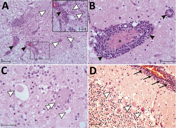
Figure 4. Tissue sections of cervical spinal cord (A), brain stem (B, C) and cerebellum (D) stained with hematoxylin and eosin from a symptomatic newly weaned pig from a farm in Hungary show the signs of stage 3 encephalomyelitis. Mononuclear perivascular cuffs with vasculitis (black arrowheads), neuronal necrosis (white arrowheads), neurophagia (white double arrowheads), multifocal microgliosis, and signs of meningitis (black arrows) are shown. Asterick (*) indicates blood vessel. Scale bars indicate 50 µm (panels A, D) or 20 µm (panel A inset; panels B, C).
Page created: November 16, 2017
Page updated: November 16, 2017
Page reviewed: November 16, 2017
The conclusions, findings, and opinions expressed by authors contributing to this journal do not necessarily reflect the official position of the U.S. Department of Health and Human Services, the Public Health Service, the Centers for Disease Control and Prevention, or the authors' affiliated institutions. Use of trade names is for identification only and does not imply endorsement by any of the groups named above.