Skip directly to site content Skip directly to page options Skip directly to A-Z link Skip directly to A-Z link Skip directly to A-Z link
Volume 23, Number 12—December 2017
Research

Outbreaks of Neuroinvasive Astrovirus Associated with Encephalomyelitis, Weakness, and Paralysis among Weaned Pigs, Hungary

Ákos Boros, Mihály Albert, Péter Pankovics, Hunor Bíró, Patricia A. Pesavento, Tung Gia Phan, Eric Delwart, and Gábor ReuterComments to Author 
Author affiliations: ÁNTSZ Regional Institute of State Public Health Service, Pécs, Hungary (A. Boros, P. Pankovics, G. Reuter); University of Pécs, Pécs (A. Boros, P. Pankovics, G. Reuter); Ceva Phylaxia Ltd., Budapest, Hungary (M. Albert); SHP Ltd., Kaposvár, Hungary (H. Bíró); University of California, Davis, California, USA (P.A. Pesavento); Blood Systems Research Institute, San Francisco, California, USA (T.G. Phan, E. Delwart); University of California, San Francisco (E. Delwart)

Main Article

Figure 5

Results of histopathologic testing of central nervous system tissues from 2 symptomatic newly weaned pigs from a farm in Hungary. Sections of the cervical spinal cord (A–E), cerebellum (F–J), and cortex (L, M) from the index animal (GD-1) and the brainstem (K) from an additional affected stage 1 animal (GD-11). A, D, F, I, L) Hematoxylin and eosin stain. Gliosis (black arrows) is multifocal within the gray matter (panels A, D) and in the molecular layers (panels F, I, L and M). Neuronal degenera

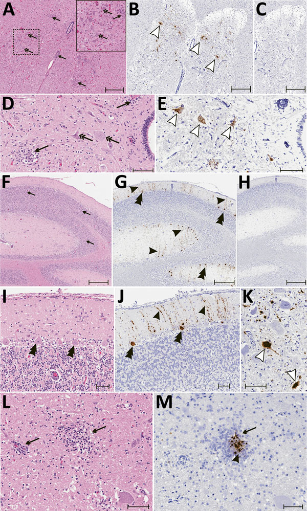
Figure 5. Results of histopathologic testing of central nervous system tissues from 2 symptomatic newly weaned pigs from a farm in Hungary. Sections of the cervical spinal cord (A–E), cerebellum (F–J), and cortex (L, M) from the index animal (GD-1) and the brain stem (K) from an additional affected stage 1 animal (GD-11). A, D, F, I, L) Hematoxylin and eosin stain. Gliosis (black arrows) is multifocal within the gray matter (panels A, D) and in the molecular layers (panels F, I, L and M). Neuronal degeneration and necrosis are evident by hypereosinophilia, angular degeneration, loss of neuronal detail, and vacuolation (double arrows in panels A, D). Some Purkinje neurons are slightly angular with mild vacuolation (double arrowheads in panel I). B, E, G, J, K, M) In situ hybridization of neuroinvasive porcine astrovirus. Hybridization of the neuroinvasive porcine astrovirus probe is restricted to neurons (white arrowheads in panels B, E, K) or limited to Purkinje neurons (double black arrowheads in panels G, J) with extension into dendritic processes that course through the molecular layer (black arrowheads in panels G, J). Hybridization of the neuroinvasive porcine astrovirus type 3 probe (black arrowhead in panel M) is present in the gliosis (black arrows in panels L, M). C, H) Using a control probe on a serial section, no hybridization is detectable. In situ hybridization. Scale bars indicate 500 µm (panels A–C, F–H) or 50 µm (panels D, E, I–M).

Main Article

Page created: November 16, 2017
Page updated: November 16, 2017
Page reviewed: November 16, 2017
The conclusions, findings, and opinions expressed by authors contributing to this journal do not necessarily reflect the official position of the U.S. Department of Health and Human Services, the Public Health Service, the Centers for Disease Control and Prevention, or the authors' affiliated institutions. Use of trade names is for identification only and does not imply endorsement by any of the groups named above.
file_external