Articles from Emerging Infectious Diseases
Volume 31, Number 9—September 2025
Perspective
Chagas Disease, an Endemic Disease in the United States
Chagas disease, caused by Trypanosoma cruzi parasites, is considered endemic to 21 countries in the Americas, excluding the United States. However, increasing evidence of T. cruzi parasites in the United States in triatomine insects, domestic animals, wildlife, and humans challenges that nonendemic label. Several triatomine species are common in the southern United States, where they transmit T. cruzi and invade human dwellings. Wildlife, captive animals, and companion animals, especially dogs, are commonly infected with T. cruzi parasites in this region and serve as reservoirs. Autochthonous human cases have been reported in 8 states, most notably in Texas. Labeling the United States as non–Chagas disease–endemic perpetuates low awareness and underreporting. Classification of Chagas disease as endemic, in particular as hypoendemic, to the United States could improve surveillance, research, and public health responses. Acknowledging the endemicity of Chagas disease in the United States is crucial for achieving global health goals.
| EID | Beatty NL, Hamer GL, Moreno-Peniche B, Mayes B, Hamer SA. Chagas Disease, an Endemic Disease in the United States. Emerg Infect Dis. 2025;31(9):1691-1697. https://doi.org/10.3201/eid3109.241700 |
|---|---|
| AMA | Beatty NL, Hamer GL, Moreno-Peniche B, et al. Chagas Disease, an Endemic Disease in the United States. Emerging Infectious Diseases. 2025;31(9):1691-1697. doi:10.3201/eid3109.241700. |
| APA | Beatty, N. L., Hamer, G. L., Moreno-Peniche, B., Mayes, B., & Hamer, S. A. (2025). Chagas Disease, an Endemic Disease in the United States. Emerging Infectious Diseases, 31(9), 1691-1697. https://doi.org/10.3201/eid3109.241700. |
Research
Sierra Colomina M, Flamant A, Le Balle G, Cohen JF, Berthomieu L, Leteurtre S, et al. Severe group A Streptococcus infection among children, France, 2022–2024. Emerg Infect Dis. 2025 Sep [date cited]. https://doi.org/10.3201/eid3109.250245
Group A Streptococcus infections have increased in Europe since September 2022. The French Pediatric Intensive Care and French Pediatric Infectious Diseases expert groups conducted a retrospective and prospective study of children who had severe group A Streptococcus infections during September 1, 2022–April 1, 2024, across 34 hospitals in France. A total of 402 pediatric patients (median age 4 [interquartile range 2–7.5] years; 42% girls, 58% boys) were enrolled. Cases were characterized by a low proportion of severe skin and soft tissue infections (16%), predominance of severe upper and lower respiratory tract infections (55%), and a 3.5% case-fatality rate. In multivariate analysis, hydrocortisone, corticosteroid, and vasopressor therapies were significantly associated with major sequelae or death. Molecular analysis revealed emm1 (73.0%) and emm12 (10.8%) strains; the M1UK clone represented 50% of emm1 strains. Clinicians, researchers, and public health authorities must collaborate to mitigate the effects of GAS on child health.
| EID | Colomina M, Flamant A, Le Balle G, Cohen JF, Berthomieu L, Leteurtre S, et al. Severe Group A Streptococcus Infection among Children, France, 2022–2024. Emerg Infect Dis. 2025;31(9):1698-1707. https://doi.org/10.3201/eid3109.250245 |
|---|---|
| AMA | Colomina M, Flamant A, Le Balle G, et al. Severe Group A Streptococcus Infection among Children, France, 2022–2024. Emerging Infectious Diseases. 2025;31(9):1698-1707. doi:10.3201/eid3109.250245. |
| APA | Colomina, M., Flamant, A., Le Balle, G., Cohen, J. F., Berthomieu, L., Leteurtre, S....Brehin, C. (2025). Severe Group A Streptococcus Infection among Children, France, 2022–2024. Emerging Infectious Diseases, 31(9), 1698-1707. https://doi.org/10.3201/eid3109.250245. |
The complexity of rickettsial serodiagnostics during acute illness has limited clinical characterization in Africa. We used archived samples from sepsis (n = 259) and acute febrile illness (n = 70) cohorts in Uganda to identify spotted fever and typhus group rickettsiae by using immunofluorescence assay and clinically validated rRNA reverse transcription PCR (RT-PCR). Among 329 participants, 10.0% had rickettsial infections (n = 33; n = 20 identified with immunofluorescence assay and n = 13 by RT-PCR). Serum rRNA RT-PCR was 75.0% (95% CI 42.8–94.5%) sensitive and 91.2% (95% CI 85.8–95.1%) specific. Thrombocytopenia was more common among patients with rickettsial infections than with other nonmalarial infections (adjusted odds ratio 3.7; p = 0.003). No participants were on a tetracycline antimicrobial drug at admission. rRNA RT-PCR is a promising diagnostic strategy for identifying acute rickettsial infections. Doxycycline should be included in empiric antimicrobial drug regimens for nonmalarial febrile illness in this region.
| EID | Blair PW, Alharthi S, Londoño AF, Wailagala A, Manabe YC, Dumler J. Rickettsioses as Underrecognized Cause of Hospitalization for Febrile Illness, Uganda. Emerg Infect Dis. 2025;31(9):1708-1717. https://doi.org/10.3201/eid3109.250479 |
|---|---|
| AMA | Blair PW, Alharthi S, Londoño AF, et al. Rickettsioses as Underrecognized Cause of Hospitalization for Febrile Illness, Uganda. Emerging Infectious Diseases. 2025;31(9):1708-1717. doi:10.3201/eid3109.250479. |
| APA | Blair, P. W., Alharthi, S., Londoño, A. F., Wailagala, A., Manabe, Y. C., & Dumler, J. (2025). Rickettsioses as Underrecognized Cause of Hospitalization for Febrile Illness, Uganda. Emerging Infectious Diseases, 31(9), 1708-1717. https://doi.org/10.3201/eid3109.250479. |
Epidemiology of Chikungunya Hospitalizations, Brazil, 2014–2024
We describe 7,421 chikungunya hospitalizations in Brazil covered by the country’s unified health system during 2014–2024. Most (43.2%) hospitalizations occurred in 2016 and 2017, reaching 0.72 (95% CI 0.69–0.76) hospitalizations/100,000 population in 2016. Hospitalizations were more frequent among persons who were female (55.8%), identifying as brown or black (63.5%), and 1–19 years of age (31.4%). Intensive care unit admissions occurred in 1.4% of cases, predominantly among children <5 and adults >85 years of age. The overall in-hospital case-fatality rate was 1.1%, which increased substantially with age, reaching 11.5% among patients >90 years of age and 14.1% among men 85–89 years of age. Patients admitted to the intensive care unit had a case-fatality rate of 21.1%. The total cost of chikungunya hospitalizations was US $560,746 (US $76.26 per patient). Our findings provide insights for surveillance of the most severe chikungunya cases.
| EID | Pedí V, Porto D, de Jesus Martins W, de França G. Epidemiology of Chikungunya Hospitalizations, Brazil, 2014–2024. Emerg Infect Dis. 2025;31(9):1718-1728. https://doi.org/10.3201/eid3109.250554 |
|---|---|
| AMA | Pedí V, Porto D, de Jesus Martins W, et al. Epidemiology of Chikungunya Hospitalizations, Brazil, 2014–2024. Emerging Infectious Diseases. 2025;31(9):1718-1728. doi:10.3201/eid3109.250554. |
| APA | Pedí, V., Porto, D., de Jesus Martins, W., & de França, G. (2025). Epidemiology of Chikungunya Hospitalizations, Brazil, 2014–2024. Emerging Infectious Diseases, 31(9), 1718-1728. https://doi.org/10.3201/eid3109.250554. |
Drivers of Crimean-Congo Hemorrhagic Fever in Natural Host and Effects of Control Measures, Bulgaria
Crimean-Congo hemorrhagic fever (CCHF) is an emerging tickborne disease and a World Health Organization priority. Although humans are accidental hosts, infection can lead to hemorrhagic fever with a high fatality rate. Domestic animals play a critical role in disease transmission, but infected animals do not show clinical signs and viremia is short; thus, CCHF virus (CCHFV) infections can remain unobserved. During 2017–2019, we conducted 2 sequential observational studies followed by a multisite randomized controlled trial to determine spatial-temporal patterns and quantify drivers for CCHFV exposure in a natural host (sheep) in a CCHF-endemic area of Bulgaria. We found high-risk areas embedded in endemic regions. Animal characteristics were not correlated with seropositivity; however, a seasonality effect was observed, suggesting sampling time was a potential confounder. Force of infection varied across farms and over time. CCHFV transmission heterogeneity among farms is driven by preventive measures used to reduce exposure to ticks.
| EID | Limon G, Tchakarova SR, Ludi A, Alexandrov T, Christova I, Petkova P, et al. Drivers of Crimean-Congo Hemorrhagic Fever in Natural Host and Effects of Control Measures, Bulgaria. Emerg Infect Dis. 2025;31(9):1738-1746. https://doi.org/10.3201/eid3109.241952 |
|---|---|
| AMA | Limon G, Tchakarova SR, Ludi A, et al. Drivers of Crimean-Congo Hemorrhagic Fever in Natural Host and Effects of Control Measures, Bulgaria. Emerging Infectious Diseases. 2025;31(9):1738-1746. doi:10.3201/eid3109.241952. |
| APA | Limon, G., Tchakarova, S. R., Ludi, A., Alexandrov, T., Christova, I., Petkova, P....Lyons, N. A. (2025). Drivers of Crimean-Congo Hemorrhagic Fever in Natural Host and Effects of Control Measures, Bulgaria. Emerging Infectious Diseases, 31(9), 1738-1746. https://doi.org/10.3201/eid3109.241952. |
Increased Incidence of Candida auris Colonization in Early COVID-19 Pandemic, Orange County, California, USA
Candida auris transmission surged in long-term acute-care hospitals (LTACHs) in Orange County, California, USA, during the COVID-19 pandemic. This study describes the effect of COVID-19 on C. auris transmission by estimating the probability of patient colonization in LTACHs across 5 epidemiologic time periods. Patients had the highest probability of developing new skin colonization during the first COVID-19 wave, with a cumulative incidence of 22.5% (95% CI 18.5–26.6) after a 30-day stay. Once the initial COVID-19 waves abated, a reduction in cumulative incidence of C. auris colonization was observed concurrently with persistent high prevalence, indicating that within-facility transmission can be reduced with proper infection prevention and control practices. Admission screenings and point prevalence surveys provided a wealth of data that guided public health recommendations and supported the objectives of both public health professionals and LTACHs for monitoring facility transmission dynamics and guiding decision making.
| EID | Dratch AH, Le M, Zahn M. Increased Incidence of Candida auris Colonization in Early COVID-19 Pandemic, Orange County, California, USA. Emerg Infect Dis. 2025;31(9):1747-1754. https://doi.org/10.3201/eid3109.241342 |
|---|---|
| AMA | Dratch AH, Le M, Zahn M. Increased Incidence of Candida auris Colonization in Early COVID-19 Pandemic, Orange County, California, USA. Emerging Infectious Diseases. 2025;31(9):1747-1754. doi:10.3201/eid3109.241342. |
| APA | Dratch, A. H., Le, M., & Zahn, M. (2025). Increased Incidence of Candida auris Colonization in Early COVID-19 Pandemic, Orange County, California, USA. Emerging Infectious Diseases, 31(9), 1747-1754. https://doi.org/10.3201/eid3109.241342. |
Differences in Lyme Disease Diagnosis among Medicaid and Medicare Beneficiaries, United States, 2016–2021
Lyme disease is the most common vectorborne disease in the United States. Evidence suggests that persons from racial and ethnic minority groups experience more severe disease. We used a claims-based algorithm on data from 16 jurisdictions with high Lyme disease incidence to identify cases among 4 populations: Medicaid beneficiaries <18 and >19 years of age, and Medicare fee-for-service beneficiaries <65 and >65 years of age. We calculated the prevalence of disseminated disease, hospitalization, and other clinical and epidemiologic parameters by race and ethnicity. We found that non-White persons were more likely than White persons to be female, hospitalized at diagnosis, diagnosed outside of primary care, diagnosed outside of the peak months for Lyme disease transmission, and have disseminated disease. Those data illustrate differences in Lyme disease by race and ethnicity and suggest possible differences across other sociodemographic characteristics. Additional prevention methods are needed to reduce differences in Lyme disease recognition and severity.
| EID | Gould L, Willis SJ, Prener CG, Duench SA, Yu H, Jodar L, et al. Differences in Lyme Disease Diagnosis among Medicaid and Medicare Beneficiaries, United States, 2016–2021. Emerg Infect Dis. 2025;31(9):1755-1763. https://doi.org/10.3201/eid3109.241653 |
|---|---|
| AMA | Gould L, Willis SJ, Prener CG, et al. Differences in Lyme Disease Diagnosis among Medicaid and Medicare Beneficiaries, United States, 2016–2021. Emerging Infectious Diseases. 2025;31(9):1755-1763. doi:10.3201/eid3109.241653. |
| APA | Gould, L., Willis, S. J., Prener, C. G., Duench, S. A., Yu, H., Jodar, L....Stark, J. H. (2025). Differences in Lyme Disease Diagnosis among Medicaid and Medicare Beneficiaries, United States, 2016–2021. Emerging Infectious Diseases, 31(9), 1755-1763. https://doi.org/10.3201/eid3109.241653. |
Theileria luwenshuni and Novel Babesia spp. Infections in Humans, Yunnan Province, China
Piroplasmid parasites such as Theileria luwenshuni protozoa pose a global threat to both animal and human health. However, human theileriosis remains underexplored compared to infections caused by Plasmodium and Babesia species parasites. We investigated potential hemoparasite infections among 1,721 persons with fever, anemia, or both in Yunnan Province, China. Molecular detection identified 13 cases positive for T. luwenshuni protozoa, of which 5 patients were further confirmed by Western blot antibody analysis. We also identified 6 babesiosis cases, 3 infections with B. microti and 3 with novel Babesia spp. Subsequent vector and host investigations in the vicinity of the index cases revealed T. luwenshuni protozoa in 1 tick and 53 livestock animals. Of note, 3.3% combined vector-host samples tested positive for genetically diverse Babesia species. Our findings highlight the endemic circulation of T. luwenshuni and Babesia spp. parasites in southwest China, underscoring their importance as emerging public health concerns.
| EID | Xiang R, Du C, Zhao Y, Luo Z, Li M, Zeng D, et al. Theileria luwenshuni and Novel Babesia spp. Infections in Humans, Yunnan Province, China. Emerg Infect Dis. 2025;31(9):1764-1773. https://doi.org/10.3201/eid3109.241919 |
|---|---|
| AMA | Xiang R, Du C, Zhao Y, et al. Theileria luwenshuni and Novel Babesia spp. Infections in Humans, Yunnan Province, China. Emerging Infectious Diseases. 2025;31(9):1764-1773. doi:10.3201/eid3109.241919. |
| APA | Xiang, R., Du, C., Zhao, Y., Luo, Z., Li, M., Zeng, D....Jiang, J. (2025). Theileria luwenshuni and Novel Babesia spp. Infections in Humans, Yunnan Province, China. Emerging Infectious Diseases, 31(9), 1764-1773. https://doi.org/10.3201/eid3109.241919. |
Detection of Multiple Nosocomial Trichosporon asahii Transmission Events via Microsatellite Typing Assay, South America
The fungus Trichosporon asahii has emerged as a cause of nosocomial infections, particularly in immunocompromised patients. Given its rising prevalence, information on its genetic diversity and transmission dynamics is urgently needed. We developed a microsatellite typing tool to investigate the genetic relatedness of T. asahii isolates. We selected 6 microsatellite markers from nanopore long-read sequencing of the T. asahii type-strain CBS 2479. We applied those markers to 111 clinical and environmental isolates; microsatellite typing showed high variability among isolates (11–37 alleles per marker) and identified 71 genotypes with strong discriminatory power (Simpson index of 0.9793). We applied the microsatellite typing method to T. asahii isolates from South America and identified multiple nosocomial transmission events from hospitals in Brazil, including clusters spanning more than a decade. The panel we developed offers high reproducibility and specificity, making it an effective tool for tracking outbreaks and determining the public health effects of T. asahii infections.
| EID | Francisco EC, Fernández NB, Carbia M, Dieleman C, Bostanaru-Iliescu A, Houbraken J, et al. Detection of Multiple Nosocomial Trichosporon asahii Transmission Events via Microsatellite Typing Assay, South America. Emerg Infect Dis. 2025;31(9):1774-1782. https://doi.org/10.3201/eid3109.241929 |
|---|---|
| AMA | Francisco EC, Fernández NB, Carbia M, et al. Detection of Multiple Nosocomial Trichosporon asahii Transmission Events via Microsatellite Typing Assay, South America. Emerging Infectious Diseases. 2025;31(9):1774-1782. doi:10.3201/eid3109.241929. |
| APA | Francisco, E. C., Fernández, N. B., Carbia, M., Dieleman, C., Bostanaru-Iliescu, A., Houbraken, J....Hagen, F. (2025). Detection of Multiple Nosocomial Trichosporon asahii Transmission Events via Microsatellite Typing Assay, South America. Emerging Infectious Diseases, 31(9), 1774-1782. https://doi.org/10.3201/eid3109.241929. |
Sporothrix brasiliensis Treatment Failure without Initial Elevated Itraconazole MICs in Felids at Border of Brazil
Cat-transmitted sporotrichosis caused by Sporothrix brasiliensis is an emerging zoonosis in Latin America. Because treatment of feline sporotrichosis is often not effective, we sought to determine whether treatment failure results from S. brasiliensis strains that have existing elevated MICs for itraconazole, the primary treatment for this disease. During 2021–2023 at the triple border region of Brazil, Paraguay, and Argentina, 108 S. brasiliensis strains were isolated from felines before antifungal treatment. The main clinical manifestation was cutaneous disseminated sporotrichosis (61%), which was the only form resulting in sporotrichosis-induced deaths (61%). We conducted antifungal susceptibility testing for 9 antifungal compounds, evaluating for both mycelial and yeast phases. MIC levels were low for most antifungal agents but were higher in the mycelial phase than in the yeast phase, especially for voriconazole and isavuconazole. We conclude that the varying clinical manifestations of sporotrichosis and large differences in mortality rates were not caused by elevated itraconazole MICs.
| EID | do Prado C, Spruijtenburg B, Razzolini E, Chiyo L, Santi C, Martins C, et al. Sporothrix brasiliensis Treatment Failure without Initial Elevated Itraconazole MICs in Felids at Border of Brazil. Emerg Infect Dis. 2025;31(9):1783-1792. https://doi.org/10.3201/eid3109.250156 |
|---|---|
| AMA | do Prado C, Spruijtenburg B, Razzolini E, et al. Sporothrix brasiliensis Treatment Failure without Initial Elevated Itraconazole MICs in Felids at Border of Brazil. Emerging Infectious Diseases. 2025;31(9):1783-1792. doi:10.3201/eid3109.250156. |
| APA | do Prado, C., Spruijtenburg, B., Razzolini, E., Chiyo, L., Santi, C., Martins, C....Queiroz-Telles, F. (2025). Sporothrix brasiliensis Treatment Failure without Initial Elevated Itraconazole MICs in Felids at Border of Brazil. Emerging Infectious Diseases, 31(9), 1783-1792. https://doi.org/10.3201/eid3109.250156. |
Insights into Infant Strongyloidiasis, Papua New Guinea
The human-infecting parasite Strongyloides fuelleborni subspecies kellyi has been reported from the island of New Guinea. We analyzed fecal DNA extracts (n = 164) from 19 infants in Papua New Guinea by using Strongyloides real-time PCR and undertook metabarcoding of cox1 and 18S rRNA hypervariable regions I and IV loci. Eight infants were infected with Strongyloides spp.; 7 were infected with S. fuelleborni subsp. fuelleborni and 1 with a Strongyloides sp. previously misattributed to S. fuelleborni subsp. kellyi. Phylogenetic and haplotyping analyses indicated S. fuelleborni in Papua New Guinea belongs to the Indochina subclade of S. fuelleborni subsp. fuelleborni and is not a unique subspecies. We report molecular evidence of S. fuelleborni subsp. fuelleborni infection in humans in the Pacific. Our findings also demonstrate the potential co-existence of an undescribed human-infecting Strongyloides sp. on the island of New Guinea, indicating a need for renewed clinical and epidemiologic investigations into infant strongyloidiasis.
| EID | Zhao H, Haidamak J, Noskova E, Ilik V, Pafčo B, Ford R, et al. Insights into Infant Strongyloidiasis, Papua New Guinea. Emerg Infect Dis. 2025;31(9):1793-1801. https://doi.org/10.3201/eid3109.241923 |
|---|---|
| AMA | Zhao H, Haidamak J, Noskova E, et al. Insights into Infant Strongyloidiasis, Papua New Guinea. Emerging Infectious Diseases. 2025;31(9):1793-1801. doi:10.3201/eid3109.241923. |
| APA | Zhao, H., Haidamak, J., Noskova, E., Ilik, V., Pafčo, B., Ford, R....Bradbury, R. S. (2025). Insights into Infant Strongyloidiasis, Papua New Guinea. Emerging Infectious Diseases, 31(9), 1793-1801. https://doi.org/10.3201/eid3109.241923. |
Dispatches
Related Melioidosis Cases with Unknown Exposure Source, Georgia, USA, 1983–2024
We identified 4 cases of presumptive autochthonous melioidosis during 1983–2024 in Georgia, USA. Epidemiologic investigation identified no recent international travel before illness; all cases were geographically linked, and 3 patients became ill after a severe weather event. Bioinformatic analyses revealed Burkholderia pseudomallei genome sequences were highly related, suggesting a shared exposure.
| EID | Brennan S, Thompson JM, Gulvik CA, Paisie TK, Elrod M, Gee JE, et al. Related Melioidosis Cases with Unknown Exposure Source, Georgia, USA, 1983–2024. Emerg Infect Dis. 2025;31(9):1802-1806. https://doi.org/10.3201/eid3109.250804 |
|---|---|
| AMA | Brennan S, Thompson JM, Gulvik CA, et al. Related Melioidosis Cases with Unknown Exposure Source, Georgia, USA, 1983–2024. Emerging Infectious Diseases. 2025;31(9):1802-1806. doi:10.3201/eid3109.250804. |
| APA | Brennan, S., Thompson, J. M., Gulvik, C. A., Paisie, T. K., Elrod, M., Gee, J. E....Gabel, J. (2025). Related Melioidosis Cases with Unknown Exposure Source, Georgia, USA, 1983–2024. Emerging Infectious Diseases, 31(9), 1802-1806. https://doi.org/10.3201/eid3109.250804. |
CYP2D6 Genotype and Primaquine Treatment in Patients with Malaria, Venezuela
We determined CYP2D6*4 and CYP2D6* genotypes and metabolizer phenotypes in 96 patients with suspected malaria in Venezuela and found intermediate or poor metabolizer phenotypes in ≈25% of cases. Nine of 44 malaria patients had Plasmodium vivax recurrence. Public health authorities should evaluate the benefits of increasing total doses of primaquine for treatment.
| EID | Pacheco C, Hernández-Acosta A, Pulido N, Ceballos Y, Saavedra D, Gómez C, et al. CYP2D6 Genotype and Primaquine Treatment in Patients with Malaria, Venezuela. Emerg Infect Dis. 2025;31(9):1807-1810. https://doi.org/10.3201/eid3109.250316 |
|---|---|
| AMA | Pacheco C, Hernández-Acosta A, Pulido N, et al. CYP2D6 Genotype and Primaquine Treatment in Patients with Malaria, Venezuela. Emerging Infectious Diseases. 2025;31(9):1807-1810. doi:10.3201/eid3109.250316. |
| APA | Pacheco, C., Hernández-Acosta, A., Pulido, N., Ceballos, Y., Saavedra, D., Gómez, C....Herrera, F. (2025). CYP2D6 Genotype and Primaquine Treatment in Patients with Malaria, Venezuela. Emerging Infectious Diseases, 31(9), 1807-1810. https://doi.org/10.3201/eid3109.250316. |
Gastric Submucosal Tumor in Patient Infected with Dioctophyme renale Roundworm, South Korea, 2024
We describe a case of a gastric submucosal tumor in a patient in South Korea infected with Dioctophyme renale roundworm. The patient had a history of consuming raw freshwater fish. Molecular and morphologic analyses confirmed D. renale Infection. Genetic testing should be used to diagnose rare parasitic infections with unusual clinical manifestations.
| EID | Kim D, Kim Y, Lee JI, Seo J, Kim D, Yun NR, et al. Gastric Submucosal Tumor in Patient Infected with Dioctophyme renale Roundworm, South Korea, 2024. Emerg Infect Dis. 2025;31(9):1811-1814. https://doi.org/10.3201/eid3109.241944 |
|---|---|
| AMA | Kim D, Kim Y, Lee JI, et al. Gastric Submucosal Tumor in Patient Infected with Dioctophyme renale Roundworm, South Korea, 2024. Emerging Infectious Diseases. 2025;31(9):1811-1814. doi:10.3201/eid3109.241944. |
| APA | Kim, D., Kim, Y., Lee, J. I., Seo, J., Kim, D., Yun, N. R....Lim, S. (2025). Gastric Submucosal Tumor in Patient Infected with Dioctophyme renale Roundworm, South Korea, 2024. Emerging Infectious Diseases, 31(9), 1811-1814. https://doi.org/10.3201/eid3109.241944. |
Rapidly Progressing Melioidosis Outbreak in City Center Zoo, Hong Kong, 2024
In October 2024, twelve primates from 4 species died of sepsis at the Hong Kong Zoological and Botanical Gardens. Postmortem examinations and microbiological analyses confirmed Burkholderia pseudomallei infection. Phylogenetic analysis revealed a clonal sequence type 46 strain with minimal variation, signifying a single source. This outbreak highlights melioidosis risk in zoo settings.
| EID | Brackman CJ, Wong I, Chan A, Pun P, Cheung D, Tse A, et al. Rapidly Progressing Melioidosis Outbreak in City Center Zoo, Hong Kong, 2024. Emerg Infect Dis. 2025;31(9):1815-1819. https://doi.org/10.3201/eid3109.250823 |
|---|---|
| AMA | Brackman CJ, Wong I, Chan A, et al. Rapidly Progressing Melioidosis Outbreak in City Center Zoo, Hong Kong, 2024. Emerging Infectious Diseases. 2025;31(9):1815-1819. doi:10.3201/eid3109.250823. |
| APA | Brackman, C. J., Wong, I., Chan, A., Pun, P., Cheung, D., Tse, A....Sit, T. (2025). Rapidly Progressing Melioidosis Outbreak in City Center Zoo, Hong Kong, 2024. Emerging Infectious Diseases, 31(9), 1815-1819. https://doi.org/10.3201/eid3109.250823. |
Genetic Characterization of Orientia tsutsugamushi, Bhutan, 2015
We performed molecular characterization of Orientia tsutsugamushi on DNA sequences from 5 patients from Bhutan with scrub typhus. In the 56 kDa gene, all isolates aligned with those from other Asia countries, including Nepal, India, Thailand, and Taiwan. High serum IgM titers correlated with PCR positivity in acutely ill patients.
| EID | Tshokey T, Stenos J, Tadepalli M, Nguyen C, Graves SR. Genetic Characterization of Orientia tsutsugamushi, Bhutan, 2015. Emerg Infect Dis. 2025;31(9):1820-1823. https://doi.org/10.3201/eid3109.241763 |
|---|---|
| AMA | Tshokey T, Stenos J, Tadepalli M, et al. Genetic Characterization of Orientia tsutsugamushi, Bhutan, 2015. Emerging Infectious Diseases. 2025;31(9):1820-1823. doi:10.3201/eid3109.241763. |
| APA | Tshokey, T., Stenos, J., Tadepalli, M., Nguyen, C., & Graves, S. R. (2025). Genetic Characterization of Orientia tsutsugamushi, Bhutan, 2015. Emerging Infectious Diseases, 31(9), 1820-1823. https://doi.org/10.3201/eid3109.241763. |
Novel Henipavirus, Salt Gully Virus, Isolated from Pteropid Bats, Australia
We describe isolation and characterization of a novel henipavirus, designated Salt Gully virus, from the urine of pteropid bats in Australia. We noted the virus to be most closely related to Angavokely virus, not reliant on ephrin receptors for cell entry, and of unknown risk for human disease.
| EID | Barr J, Caruso S, Edwards SJ, Todd S, Smith I, Tachedjian M, et al. Novel Henipavirus, Salt Gully Virus, Isolated from Pteropid Bats, Australia. Emerg Infect Dis. 2025;31(9):1824-1828. https://doi.org/10.3201/eid3109.250470 |
|---|---|
| AMA | Barr J, Caruso S, Edwards SJ, et al. Novel Henipavirus, Salt Gully Virus, Isolated from Pteropid Bats, Australia. Emerging Infectious Diseases. 2025;31(9):1824-1828. doi:10.3201/eid3109.250470. |
| APA | Barr, J., Caruso, S., Edwards, S. J., Todd, S., Smith, I., Tachedjian, M....Marsh, G. A. (2025). Novel Henipavirus, Salt Gully Virus, Isolated from Pteropid Bats, Australia. Emerging Infectious Diseases, 31(9), 1824-1828. https://doi.org/10.3201/eid3109.250470. |
Modeling Case Burden and Duration of Sudan Ebola Virus Disease Outbreak in Uganda, 2022
In 2022, a Sudan Ebola virus outbreak was confirmed in Uganda. Within 1 month of the outbreak’s onset, we developed an individual-based modeling platform to estimate the unfolding outbreak’s burden of cases and deaths, as well as its duration, using different scenarios. Modeled projections were within the range of observed cases.
| EID | Bisanzio D, Bosa H, Bakamutumaho B, Nasimiyu C, Atwine D, Kyabayinze D, et al. Modeling Case Burden and Duration of Sudan Ebola Virus Disease Outbreak in Uganda, 2022. Emerg Infect Dis. 2025;31(9):1829-1832. https://doi.org/10.3201/eid3109.241545 |
|---|---|
| AMA | Bisanzio D, Bosa H, Bakamutumaho B, et al. Modeling Case Burden and Duration of Sudan Ebola Virus Disease Outbreak in Uganda, 2022. Emerging Infectious Diseases. 2025;31(9):1829-1832. doi:10.3201/eid3109.241545. |
| APA | Bisanzio, D., Bosa, H., Bakamutumaho, B., Nasimiyu, C., Atwine, D., Kyabayinze, D....Reithinger, R. (2025). Modeling Case Burden and Duration of Sudan Ebola Virus Disease Outbreak in Uganda, 2022. Emerging Infectious Diseases, 31(9), 1829-1832. https://doi.org/10.3201/eid3109.241545. |
Detection of Rat Lungworm (Angiostrongylus cantonensis) in Rats and Gastropods, Italy
The emerging zoonotic nematode Angiostrongylus cantonensis causes severe neural angiostrongyliasis in both humans and animals. The parasite has been reported in Spain. We detected A. cantonensis in rats and gastropods from the Campania region, southern Italy, demonstrating its broad distribution on the southern coast of Europe.
| EID | Pandian D, Šipková A, Scarcelli S, Sgroi G, Kačmaříková J, Buono F, et al. Detection of Rat Lungworm (Angiostrongylus cantonensis) in Rats and Gastropods, Italy. Emerg Infect Dis. 2025;31(9):1833-1837. https://doi.org/10.3201/eid3109.250648 |
|---|---|
| AMA | Pandian D, Šipková A, Scarcelli S, et al. Detection of Rat Lungworm (Angiostrongylus cantonensis) in Rats and Gastropods, Italy. Emerging Infectious Diseases. 2025;31(9):1833-1837. doi:10.3201/eid3109.250648. |
| APA | Pandian, D., Šipková, A., Scarcelli, S., Sgroi, G., Kačmaříková, J., Buono, F....Modrý, D. (2025). Detection of Rat Lungworm (Angiostrongylus cantonensis) in Rats and Gastropods, Italy. Emerging Infectious Diseases, 31(9), 1833-1837. https://doi.org/10.3201/eid3109.250648. |
Emergence of Autochthonous Leishmania (Mundinia) martiniquensis Infections in Horses, Czech Republic and Austria, 2019–2023
We report 4 cases of equine cutaneous leishmaniasis caused by Leishmania martiniquensis in Czech Republic and Austria, outside the known endemic range of leishmaniases. The parasite should be considered as a potential cause of cutaneous lesions in horses; the risk for zoonotic transmission to immunocompromised humans is anticipated throughout central Europe.
| EID | Modrý D, Hainisch EK, Fuehrer H, Kniha E, Unterköfler M, Sádlová J, et al. Emergence of Autochthonous Leishmania (Mundinia) martiniquensis Infections in Horses, Czech Republic and Austria, 2019–2023. Emerg Infect Dis. 2025;31(9):1838-1842. https://doi.org/10.3201/eid3109.250254 |
|---|---|
| AMA | Modrý D, Hainisch EK, Fuehrer H, et al. Emergence of Autochthonous Leishmania (Mundinia) martiniquensis Infections in Horses, Czech Republic and Austria, 2019–2023. Emerging Infectious Diseases. 2025;31(9):1838-1842. doi:10.3201/eid3109.250254. |
| APA | Modrý, D., Hainisch, E. K., Fuehrer, H., Kniha, E., Unterköfler, M., Sádlová, J....Votýpka, J. (2025). Emergence of Autochthonous Leishmania (Mundinia) martiniquensis Infections in Horses, Czech Republic and Austria, 2019–2023. Emerging Infectious Diseases, 31(9), 1838-1842. https://doi.org/10.3201/eid3109.250254. |
Imported Malaria and Congenital Acquisition in Infant, Portugal, 2024
Plasmodium falciparum infection was diagnosed in a 3-month-old baby in Portugal by optical microscopy. The mother had had malaria in Angola 13 months earlier, before she emigrated to Portugal. She remained asymptomatic throughout and after pregnancy. We confirmed the diagnosis of an imported malaria case and congenital transmission using molecular techniques.
| EID | Lopes I, Dias J, Das Neves E, Morais M, Santos-Reis A, Garcia AM, et al. Imported Malaria and Congenital Acquisition in Infant, Portugal, 2024. Emerg Infect Dis. 2025;31(9):1843-1846. https://doi.org/10.3201/eid3109.250536 |
|---|---|
| AMA | Lopes I, Dias J, Das Neves E, et al. Imported Malaria and Congenital Acquisition in Infant, Portugal, 2024. Emerging Infectious Diseases. 2025;31(9):1843-1846. doi:10.3201/eid3109.250536. |
| APA | Lopes, I., Dias, J., Das Neves, E., Morais, M., Santos-Reis, A., Garcia, A. M....Lopes, D. (2025). Imported Malaria and Congenital Acquisition in Infant, Portugal, 2024. Emerging Infectious Diseases, 31(9), 1843-1846. https://doi.org/10.3201/eid3109.250536. |
Research Letters
Monkeypox Virus Clade IIa Infections, Liberia, 2023–2024
We performed monkeypox virus genome sequencing on clinical samples from Liberia, yielding 5 clade IIa genomes. Our analysis found no evidence of sustained human-to-human transmission, suggesting independent zoonotic spillovers from a diverse viral lineage. Public health officials should continue monitoring and sequencing efforts to identify emerging monkeypox virus lineages.
| EID | Nyan D, Berry I, Shobayo BI, Mehta M, Worwa G, Hirsch S, et al. Monkeypox Virus Clade IIa Infections, Liberia, 2023–2024. Emerg Infect Dis. 2025;31(9):1848-1851. https://doi.org/10.3201/eid3109.250271 |
|---|---|
| AMA | Nyan D, Berry I, Shobayo BI, et al. Monkeypox Virus Clade IIa Infections, Liberia, 2023–2024. Emerging Infectious Diseases. 2025;31(9):1848-1851. doi:10.3201/eid3109.250271. |
| APA | Nyan, D., Berry, I., Shobayo, B. I., Mehta, M., Worwa, G., Hirsch, S....Zufan, S. E. (2025). Monkeypox Virus Clade IIa Infections, Liberia, 2023–2024. Emerging Infectious Diseases, 31(9), 1848-1851. https://doi.org/10.3201/eid3109.250271. |
Detection of Rat Lungworms in Invasive Mollusks, Georgia, USA, 2024
The rat lungworm, Angiostrongylus cantonensis, is an invasive, zoonotic parasite that can cause severe disease in humans. We collected A. cantonensis larvae from 2 host species, invasive apple and mystery snails, from bodies of water in Georgia, USA. Recreational water users should avoid ingesting potentially infected hosts, aquatic vegetation, and water.
| EID | Achatz TJ, Chun CH, Young MA, Page J, Rowe M, Cooper C, et al. Detection of Rat Lungworms in Invasive Mollusks, Georgia, USA, 2024. Emerg Infect Dis. 2025;31(9):1852-1854. https://doi.org/10.3201/eid3109.250133 |
|---|---|
| AMA | Achatz TJ, Chun CH, Young MA, et al. Detection of Rat Lungworms in Invasive Mollusks, Georgia, USA, 2024. Emerging Infectious Diseases. 2025;31(9):1852-1854. doi:10.3201/eid3109.250133. |
| APA | Achatz, T. J., Chun, C. H., Young, M. A., Page, J., Rowe, M., Cooper, C....Tkach, V. V. (2025). Detection of Rat Lungworms in Invasive Mollusks, Georgia, USA, 2024. Emerging Infectious Diseases, 31(9), 1852-1854. https://doi.org/10.3201/eid3109.250133. |
Characterization of Emerging Human Dirofilaria repens Infections, Estonia, 2023
Mosquitoborne diseases are a growing threat to public health worldwide. Human dirofilariasis, caused by the nematode Dirofilaria repens and transmitted by mosquitoes from various genera, has recently expanded into new areas of Europe. In this article, we report molecularly confirmed autochthonous human D. repens infections in Estonia.
| EID | Nõupuu K, Mõtsküla M, Pulges R, Pauklin M, Saarma U. Characterization of Emerging Human Dirofilaria repens Infections, Estonia, 2023. Emerg Infect Dis. 2025;31(9):1854-1857. https://doi.org/10.3201/eid3109.241890 |
|---|---|
| AMA | Nõupuu K, Mõtsküla M, Pulges R, et al. Characterization of Emerging Human Dirofilaria repens Infections, Estonia, 2023. Emerging Infectious Diseases. 2025;31(9):1854-1857. doi:10.3201/eid3109.241890. |
| APA | Nõupuu, K., Mõtsküla, M., Pulges, R., Pauklin, M., & Saarma, U. (2025). Characterization of Emerging Human Dirofilaria repens Infections, Estonia, 2023. Emerging Infectious Diseases, 31(9), 1854-1857. https://doi.org/10.3201/eid3109.241890. |
Zoonotic Rat Lungworm Angiostrongylus cantonensis in Black Rats, Houston, Texas, 2024
The Angiostrongylus cantonensis rat lungworm is a zoonotic nematode that infects several rat species. This nematode causes eosinophilic meningitis and meningoencephalitis in humans and other accidental hosts. We found a 20% prevalence of A. cantonensis lungworms in black rats from a zoo facility in Houston, Texas, USA.
| EID | Sierra DA, Sanders TL, Edwards EE, Molter CM, Verocai GG. Zoonotic Rat Lungworm Angiostrongylus cantonensis in Black Rats, Houston, Texas, 2024. Emerg Infect Dis. 2025;31(9):1857-1860. https://doi.org/10.3201/eid3109.251710 |
|---|---|
| AMA | Sierra DA, Sanders TL, Edwards EE, et al. Zoonotic Rat Lungworm Angiostrongylus cantonensis in Black Rats, Houston, Texas, 2024. Emerging Infectious Diseases. 2025;31(9):1857-1860. doi:10.3201/eid3109.251710. |
| APA | Sierra, D. A., Sanders, T. L., Edwards, E. E., Molter, C. M., & Verocai, G. G. (2025). Zoonotic Rat Lungworm Angiostrongylus cantonensis in Black Rats, Houston, Texas, 2024. Emerging Infectious Diseases, 31(9), 1857-1860. https://doi.org/10.3201/eid3109.251710. |
Human Babesiosis Caused by Babesia venatorum, Russia, 2024
We report a case of acute babesiosis in a splenectomized 63-year-old man in Siberia, Russia. We confirmed the causative agent, Babesia venatorum, by PCR. Our study demonstrated a change in the structure of the parasite population, from single parasite invasion of erythrocytes to multioccupancy, without an increase in parasitemia level.
| EID | Zelya OP, Kukina IV, Karan LS, Krasilovskaya EA, Garin VV. Human Babesiosis Caused by Babesia venatorum, Russia, 2024. Emerg Infect Dis. 2025;31(9):1860-1862. https://doi.org/10.3201/eid3109.250319 |
|---|---|
| AMA | Zelya OP, Kukina IV, Karan LS, et al. Human Babesiosis Caused by Babesia venatorum, Russia, 2024. Emerging Infectious Diseases. 2025;31(9):1860-1862. doi:10.3201/eid3109.250319. |
| APA | Zelya, O. P., Kukina, I. V., Karan, L. S., Krasilovskaya, E. A., & Garin, V. V. (2025). Human Babesiosis Caused by Babesia venatorum, Russia, 2024. Emerging Infectious Diseases, 31(9), 1860-1862. https://doi.org/10.3201/eid3109.250319. |
Linezolid and Meropenem for Nocardia otitidiscaviarum Actinomycetoma, India
Treatment of actinomycotic mycetoma with joint involvement is challenging. We present a patient in India with actinomycotic mycetoma who reached complete cure and remission after linezolid and meropenem treatment with a 2-year posttreatment follow-up. Clinicians should use novel drug regimens based on subspecies variations of Nocardia and regional drug susceptibility patterns to guide therapy.
| EID | Sardana K, Sharath S, Sachdeva S, Das S, Rai G, Singh P. Linezolid and Meropenem for Nocardia otitidiscaviarum Actinomycetoma, India. Emerg Infect Dis. 2025;31(9):1863-1865. https://doi.org/10.3201/eid3109.250514 |
|---|---|
| AMA | Sardana K, Sharath S, Sachdeva S, et al. Linezolid and Meropenem for Nocardia otitidiscaviarum Actinomycetoma, India. Emerging Infectious Diseases. 2025;31(9):1863-1865. doi:10.3201/eid3109.250514. |
| APA | Sardana, K., Sharath, S., Sachdeva, S., Das, S., Rai, G., & Singh, P. (2025). Linezolid and Meropenem for Nocardia otitidiscaviarum Actinomycetoma, India. Emerging Infectious Diseases, 31(9), 1863-1865. https://doi.org/10.3201/eid3109.250514. |
Subarachnoid Neurocysticercosis Caused by Larval-Stage Taenia crassiceps Tapeworm, Slovenia
We present a case of subarachnoid neurocysticercosis caused by Taenia crassiceps in an elderly woman in Slovenia with no underlying disease or immunosuppressive treatment. The parasite was identified by 12S rDNA PCR and sequencing. Despite prolonged therapy with albendazole and praziquantel, the disease recurred after treatment was discontinued.
| EID | Šoba B, Kolar S, Gačnik A, Radež M, Petrijan T, Marko J. Subarachnoid Neurocysticercosis Caused by Larval-Stage Taenia crassiceps Tapeworm, Slovenia. Emerg Infect Dis. 2025;31(9):1865-1868. https://doi.org/10.3201/eid3109.250014 |
|---|---|
| AMA | Šoba B, Kolar S, Gačnik A, et al. Subarachnoid Neurocysticercosis Caused by Larval-Stage Taenia crassiceps Tapeworm, Slovenia. Emerging Infectious Diseases. 2025;31(9):1865-1868. doi:10.3201/eid3109.250014. |
| APA | Šoba, B., Kolar, S., Gačnik, A., Radež, M., Petrijan, T., & Marko, J. (2025). Subarachnoid Neurocysticercosis Caused by Larval-Stage Taenia crassiceps Tapeworm, Slovenia. Emerging Infectious Diseases, 31(9), 1865-1868. https://doi.org/10.3201/eid3109.250014. |
Pediatric Case Report and Overview of Autochthonous Tick-Borne Encephalitis, Belgium
Prevalence of tick-borne encephalitis (TBE) is increasing in much of Europe. In May 2024, an autochthonous pediatric case of TBE was diagnosed in a 6-year-old girl in Belgium. Clinicians should recognize the symptoms and signs of TBE infections and consider this disease in patients with unexplained neurologic symptoms, regardless of travel history.
| EID | De Langhe J, Sourbron J, Van Herreweghe R, van Esbroeck M, Vercauteren K, de Block T, et al. Pediatric Case Report and Overview of Autochthonous Tick-Borne Encephalitis, Belgium. Emerg Infect Dis. 2025;31(9):1868-1870. https://doi.org/10.3201/eid3109.250093 |
|---|---|
| AMA | De Langhe J, Sourbron J, Van Herreweghe R, et al. Pediatric Case Report and Overview of Autochthonous Tick-Borne Encephalitis, Belgium. Emerging Infectious Diseases. 2025;31(9):1868-1870. doi:10.3201/eid3109.250093. |
| APA | De Langhe, J., Sourbron, J., Van Herreweghe, R., van Esbroeck, M., Vercauteren, K., de Block, T....Van Acker, J. (2025). Pediatric Case Report and Overview of Autochthonous Tick-Borne Encephalitis, Belgium. Emerging Infectious Diseases, 31(9), 1868-1870. https://doi.org/10.3201/eid3109.250093. |
New World Screwworm Infestation in Wild Mountain Tapirs, Central Andes Mountains, Colombia
We describe New World screwworm (Cochliomyia hominivorax) infestation in 2 injured mountain tapirs (Tapirus pinchaque) from a protected area in the Central Andes, Colombia. Screwworms were not a known threat to mountain tapirs. Community outreach is needed to raise awareness on effects of this parasite on humans, domestic animals, and wildlife.
| EID | Cepeda-Duque J, Cano-González L, Elejalde G, Mantilla J, Álvarez-Arellano D, Gómez-Salazar J, et al. New World Screwworm Infestation in Wild Mountain Tapirs, Central Andes Mountains, Colombia. Emerg Infect Dis. 2025;31(9):1871-1874. https://doi.org/10.3201/eid3109.250339 |
|---|---|
| AMA | Cepeda-Duque J, Cano-González L, Elejalde G, et al. New World Screwworm Infestation in Wild Mountain Tapirs, Central Andes Mountains, Colombia. Emerging Infectious Diseases. 2025;31(9):1871-1874. doi:10.3201/eid3109.250339. |
| APA | Cepeda-Duque, J., Cano-González, L., Elejalde, G., Mantilla, J., Álvarez-Arellano, D., Gómez-Salazar, J....Vélez, J. (2025). New World Screwworm Infestation in Wild Mountain Tapirs, Central Andes Mountains, Colombia. Emerging Infectious Diseases, 31(9), 1871-1874. https://doi.org/10.3201/eid3109.250339. |
Letters
Nosocomial Transmission of Plasmodium falciparum Malaria, Spain, 2024
| EID | Gómez Perales JL, Mendoza A, Amares M. Nosocomial Transmission of Plasmodium falciparum Malaria, Spain, 2024. Emerg Infect Dis. 2025;31(9):1874-1875. https://doi.org/10.3201/eid3109.250920 |
|---|---|
| AMA | Gómez Perales JL, Mendoza A, Amares M. Nosocomial Transmission of Plasmodium falciparum Malaria, Spain, 2024. Emerging Infectious Diseases. 2025;31(9):1874-1875. doi:10.3201/eid3109.250920. |
| APA | Gómez Perales, J. L., Mendoza, A., & Amares, M. (2025). Nosocomial Transmission of Plasmodium falciparum Malaria, Spain, 2024. Emerging Infectious Diseases, 31(9), 1874-1875. https://doi.org/10.3201/eid3109.250920. |
Nosocomial Transmission of Plasmodium falciparum Malaria, Spain, 2024 (Response)
| EID | Romero M, de Pipaón M, Amuedo M, Rubio Muñoz JM, Jiménez-Hoyuela JM, Cisneros JM. Nosocomial Transmission of Plasmodium falciparum Malaria, Spain, 2024 (Response). Emerg Infect Dis. 2025;31(9):1875. https://doi.org/10.3201/eid3109.251210 |
|---|---|
| AMA | Romero M, de Pipaón M, Amuedo M, et al. Nosocomial Transmission of Plasmodium falciparum Malaria, Spain, 2024 (Response). Emerging Infectious Diseases. 2025;31(9):1875. doi:10.3201/eid3109.251210. |
| APA | Romero, M., de Pipaón, M., Amuedo, M., Rubio Muñoz, J. M., Jiménez-Hoyuela, J. M., & Cisneros, J. M. (2025). Nosocomial Transmission of Plasmodium falciparum Malaria, Spain, 2024 (Response). Emerging Infectious Diseases, 31(9), 1875. https://doi.org/10.3201/eid3109.251210. |
Etymologia
Apicoplast [ā′-pik-ō-plast]
| EID | Shankar H, Shahar M, Florentin A. Apicoplast [ā′-pik-ō-plast]. Emerg Infect Dis. 2025;31(9):1847. https://doi.org/10.3201/eid3109.241446 |
|---|---|
| AMA | Shankar H, Shahar M, Florentin A. Apicoplast [ā′-pik-ō-plast]. Emerging Infectious Diseases. 2025;31(9):1847. doi:10.3201/eid3109.241446. |
| APA | Shankar, H., Shahar, M., & Florentin, A. (2025). Apicoplast [ā′-pik-ō-plast]. Emerging Infectious Diseases, 31(9), 1847. https://doi.org/10.3201/eid3109.241446. |
About the Cover
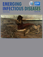
While Man Deliberates, Malaria Rules
| EID | Freedman DO. While Man Deliberates, Malaria Rules. Emerg Infect Dis. 2025;31(9):1876-1878. https://doi.org/10.3201/eid3109.ac3109 |
|---|---|
| AMA | Freedman DO. While Man Deliberates, Malaria Rules. Emerging Infectious Diseases. 2025;31(9):1876-1878. doi:10.3201/eid3109.ac3109. |
| APA | Freedman, D. O. (2025). While Man Deliberates, Malaria Rules. Emerging Infectious Diseases, 31(9), 1876-1878. https://doi.org/10.3201/eid3109.ac3109. |


